Lesiones hiperecogénicas de la mama:
¿nos quedamos tranquilos?
Palabras clave:
Lesiones hiperecogénicas, mama, poster, seramResumen
Objetivos Docentes
Mostrar el espectro de hallazgos mamarios hiperecogénicos, su correlación con otras técnicas diagnósticas y aportar las herramientas necesarias para su diagnóstico diferencial.
Revisión del tema
La ecografía mamaria es el estudio indicado en las lesiones palpables y las masas observadas en la mamografía, así como la técnica inicial en mujeres muy jóvenes para evitar la irradiación. El estudio de las características ecográficas de una lesión es muy útil para determinar su probabilidad de malignidad, la cual indicará la necesidad de estudio histológico.
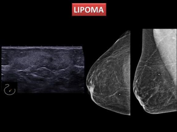
Se define como hiperecogénica aquella lesión mamaria cuya ecogenicidad sea superior a la de la grasa mamaria o igual que la del tejido fibroglandular adyacente. Estas lesiones son menos frecuentes que las hipoecogénicas, con una prevalencia según algunos estudios del 0,6 - 5,6%, y además están clásicamente asociadas a benignidad.
Descargas
Citas
Ganau , S, Tortajada , L, Escribano,F, F.J. Andreu, and M. Sentís. “Fat Necrosis.” Mammography - Recent Advances, 2012. http://www.intechopen.com/articles/show/title/fat-necrosis.
Ganau, Sergi, Lidia Tortajada, Fernanda Escribano, Xavier Andreu, and Melcior Sentís. “The Great Mimicker: Fat Necrosis of the Breast--Magnetic Resonance Mammography Approach.” Current Problems in Diagnostic Radiology 38, no. 4 (August 2009): 189–97. doi:10.1067/j.cpradiol.2009.01.001.
Stavros, A. Thomas., Cynthia L. Rapp, and Steve H. Parker. Breast Ultrasound. Philadelphia: Lippincott Williams & Wilkins, 2004. Print.
Nachiko Uchiyama and Marcelo Zanchetta do Nascimento, ed. Mammography - Recent Advances. InTech, March, 2012, 2012. http://www.intechopen.com/books/mammography-recent-advances
Radiology AC. Breast Imaging Reporting and Data System: ACR BI-RADS®. BreastImaging Atlas. 5 th Ed. 2013.
Gao Y, Slanetz MMPJ, L MPHR. Echogenic Breast Masses at US?: To Biopsy or Not to Biopsy? RadioGraphics2013:419-435.
Linda A, Lorenzon M, Furlan A, et al. Hyperechoic Lesions of the Breast: Not Always Benign. AJR 2011; 196:1219–1224.
Adrada B. Wu Yun, Yang W. Hyperechoic lesions of the breast: Radiologic-histopathologic correlation. AJR 2013; 200:518–530.
JM Sabaté, M Clotet el al. Radiologic Evaluation of Breast Disorders Related to Pregnancy and Lactation. Radiographics 2007 Oct;27 Suppl 1:S101-24. doi: 10.1148/rg.27si075505.
Irshad A et al. Rare Breast Lesions: Correlation of Imaging and Histologic Features with WHO Classification. Radiographics 2008;28(5):1399-414. doi: 10.1148/rg.285075743.
W. W. M. Lam et al. Sonographic Appearance of Mucinous Carcinoma of the Breast. AJR 2004 Apr;182(4):1069-74.
Darnell, A, X Gallardo, M Sentis, E Castañer, E Fernandez, and M Villajos. “Primary Lymphoma of the Breast: MR Imaging Features. A Case Report.” Magnetic Resonance Imaging 17, no. 3 (April 1999): 479–82. doi:10195594.
Quiles, Ana M., Lidia Tortajada, Melcior Sentís, Maite Villajos, Anna Darnell, and Xavier Andreu. “Linfoma de Mama: Hallazgos Por Resonancia Magnética Con Correlación Mamográfica Y Ecográfica.” Radiología 47, no. 1 (January 2005): 13–21. doi:10.1016/S0033-8338(05)72791-0.
Gómez, A., J. M. Mata, L. Donoso, and A. Rams. “Galactocele: Three Distinctive Radiographic Appearances.” Radiology 158, no. 1 (January 1986): 43–44.
doi:10.1148/radiology.158.1.3940395.
Bermúdez, P., and others. “Adenoma de La Lactancia: Diagnóstico Diferencial de Las Lesiones Palpables Durante El Embarazo Y La Lactancia.” Radiología 46, no. 05 (2004): 320.Jennifer A et al.. Unusual Breast Cancers: Useful Clues to Expanding the DifferentialDiagnosis. Radiology: 2007;242:3.
Wei-Hsin Yuan. Isolated panniculitis with vasculitis of the male breast suspicious for malignancy on CT and ultrasound: a case report and literature review. SpringerPlus 2014, 3:642.


