Hallazgos diagnósticos de las ictericias obstructivas no litiásicas
Palabras clave:
poster, seram, ictericia, obstructiva, no lisiatica, diagnósticoResumen
Objetivos Docentes
Manejar el amplio espectro de patología obstructiva (no litiásica) del arbol biliar.
Conocer los protocolos, técnicas de imagen adecuadas y la interpretación de las mismas (analizando hallazgos típicos/atípicos) para su correcto diagnostico.
Revisión del tema
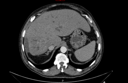
El estudio de la enfermedad del tracto biliar es un problema radiológico común. Inicialmente necesitamos determinar la presencia o ausencia de obstrucción y si se confirma definir el nivel y la causa.
Gracias a los avances en la última década en ecografía, TAC y fundamentalmente RM (que ha experimentado significativos progresos) se ha mejorado considerablemente nuestra capacidad para evaluar la vía biliar.
Descargas
Citas
Wael E. A. Saad, MBBCh, and Daniel Ginat, MD: Computed Tomography and Magnetic Resonance Cholangiography. Tech Vasc Interventional Rad 11:7489 © 2008 Elsevier Inc.
Richard M. Gore, MDa,*, Vahid Yaghmai, MDa, Geraldine M. Newmark, MDa, Jonathan W. Berlin, MDa, Frank H. Miller, MDb . Imaging benign and malignant disease of the gallbladder. Radiol Clin N Am 40 (2002) 1307– 1323
Richard L. Baron, MDa,*, Mitchell E. Tublin, MDb, Mark S. Peterson, MDb. Imaging the spectrum of biliary tract disease. Radiol Clin N Am 40 (2002) 1325– 1354
Benjamin L. Yam, MD, Evan S. Siegelman, MD. MR Imaging of the Biliary System
Neel B. Patel, MD • Aytekin Oto, MD • Stephen Thomas, MD. Multidetector CT of Emergent Biliary Pathologic
Conditions. Radiographics. See pp 2117–2124.
Eriko Maeda • Masaaki Akahane • Naoki Yoshioka • Hidemasa Takao •Izuru Matsuda • Kouhei Kamiya • Kenji Hirano • Minoru Tada • Hiroshi Ohtsu • Noriyoshi Fukushima • Kuni Ohtomo. Comparison of CT findings of biliary tract changes with autoimmune pancreatitis and extrahepatic bile duct cholangiocarcinoma. Jpn J Radiol (2012) 30:227–234 DOI 10.1007/s1160401100356
Oikarinen H. Diagnostic imaging of carcinomas of the gallbladder and the bile ducts. Acta Radiol 2006;47:345–358.
FengBo Wang,1 JianMing Ni,1 ZhuiYang Zhang,1 Lei Zhang,1 WenJuan Wu,1Dong Wang,1 Yuan Ji,2 Lei Gong3.Differential diagnosis of periampullarycarcinomas: comparison of CT with negative contrast CT cholangiopancreatography versus MRI with MR cholangiopancreatography. Abdom Imaging (2014) 39:506–517 DOI: 10.1007/s0026101400851
Bin Li & Lei Zhang & ZhuiYang Zhang & JianMing Ni &Fengqi Lu & WenJuan Wu & Chunjuan Jiang. Differentiation of noncalculous periampullary obstruction: comparison of CT with negativecontrast CT cholangiopancreatography versus MRI with MRcholangiopancreatography. Eur Radiol (2015) 25:391–401 DOI 10.1007/s0033001434304
K. Rayapudi P. Gholami M. Olyaee Kansas University Medical Center, Kansas City, Kans., USA Mirizzi Syndrome with Endoscopic Ultrasound Image. Rep Gastroenterol 2013;7:202–207
Ann S. Fulcher, MD*, Mary Ann Turner, MD. MR cholangiopancreatography. Radiol Clin N Am 40 (2002) 1363– 1376
Tomofumi Motohara, MDa, Richard C. Semelka, MDa,*, Till R. Bader, MDb. MR cholangiopancreatography. Radiol Clin
N Am 41 (2003) 89– 96
Mark S. Talamonti, MDa,*, Woody Denham, MDb.. Staging and surgical management of pancreatic and biliary cancer and inflammation Radiol Clin N Am 40 (2002) 1397– 1410.