Patología del hueco poplíteo en Urgencias.
Más allá de la TVP.
Palabras clave:
hueco poplíteo, TVP, poster, seramResumen
Objetivos Docentes
1.Mostrar las distintas patologías que pueden asentar en el hueco poplíteo.
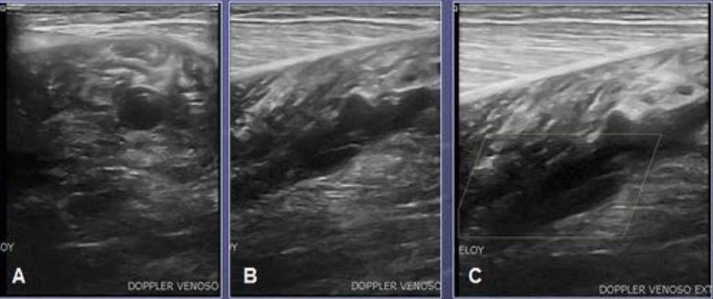
2.Describir las características ecográficas de cada una de ellas que nos van a permitir realizar un diagnóstico preciso.
Revisión del tema
Diariamente recibimos peticiones de Ecografía doppler venosa desde el Servicio de Urgencias para el estudio de pacientes con dolor en hueco poplíteo y pantorrilla. Aunque generalmente la sospecha clínica es de trombosis venosa profunda, dicho diagnóstico se confirma ecográficamente en menos del 50% de
los casos.
Existen una serie de entidades que clínica y analíticamente pueden simular una trombosis venosa: celulitis, quiste de Baker complicado, patología musculoesquelética (rotura fibrilar, hematomas), aneurisma de arteria poplítea,…
A continuación se describen las características ecográficas de estas patologías que nos van a permitir su diagnóstico una vez descartada la TVP.
Descargas
Citas
Borgstede JP, Glagett GE. Types, frequency, and significance of alternative diagnoses found during duplex Doppler venous examinations of the lower
extremities. Ultrasound Med 1992 Mar; 11(3):85-9.
Fernández-Cantón G, López Vidaur I, Muñoz F, Antoñana MA, Uresandi F, Calonge J. Diagnostic utility of color Doppler ultrasound in lower limb deep
vein thrombosis in patients with clinical suspicion of pulmonary thromboembolism. Eur J Radiol. 1994 Nov; 19(1):50-5.
Theodorou SJ, Theodorou DJ, Kakitsubata Y. Sonography and venography of the lower extremities for diagnosing deep vein thrombosis in symptomatic patients. Clin Imaging. 2003 May-Jun; 27(3):180-3.
Blumenberg RM, Barton E, Gelfand ML, Skudder P, Brennan J. Occult deep venous thrombosis complicating superficial thrombophlebitis. J Vasc Surg. 1998 Feb; 27(2):338-43.
Ward EE, Jacobson JA, Fessell DP, Hayes CW, van Holsbeeck M. Sonographic detection of Baker´s cysts: comparison with MR imaging. AJR Am J Roentgenol. 2001 Feb; 176 (2):373-80.
Handy JR. Popliteal cysts in adults: a review. Semin Arthritis Rheum. 2001 Oct;31(2):108-18.
Fang CS, McCarthy CL, McNally EG. Intramuscular dissection of Baker's cysts: report on three cases. Skeletal Radiol. 2004 Jun;33(6):367-71.
Prescott SM, Pearl JE, Tikoff G. “Pseudo- pseudothrombophlebitis”. Rupture of popliteal cyst with deep venous thrombosis. New J Engl Med 1978;299:1192-3.
Jamadar DA, Jacobson JA, Theisen SE, Marcantonio DR, Fessell DP, et al. Sonography or the painful Calf: Differential Considerations. AJR 2002; 179:709-716.
D Kane, P V Balint, R Gibney, B Bresnihan, R D Sturrock. Differential diagnosis of calf pain with musculoskeletal ultrasound imaging. Ann Rheum Dis 2004;63:11–14.
Borgstede JP, Clagett GE. Types, frequency, and significance of alternative diagnoses found during duplex Doppler venous examinations of the lower extremities. Ultrasound Med 1992 Mar; 11(3):85-9.
Perry Meier et al. Nonvascular pathology identified by duplex scanning during testing for venous thrombosis in the edematous and/or painful lower extremity JVT 1999; 23:197–200.
Loyer EM, DuBrow RA, David acl, Coan JD, Eftekari F. Imaging of Superficial Soft-Tissue Infections. Sonographics Findings in Cases of Cellulitis and Abscess. AJR 1996; 166: 149-152.
Imigo F, Fonfach C, Massri D, Sánchez G, Sánchez A. Aneurisma de arteria poplítea. Cuad Cir 2009; 23: 39-43.
Figueroa G, Pereira M, Campos A, Moreno JP, Rivera N, et al. Aneurisma arteria poplítea. Rev Chil Cir 2014; 66(5): 486-88.
Lee JC, Healy J. Sonography of Lower Limb Muscle Injury. AJR 2004; 182:341-351.
Toscano F, Iuliano GP, Grassia M, Bardascino L. The "pedrada" ("coup-defouet") syndrome. A possibility to be considered in acute leg pain. Minerva Chir 1997 Apr; 52(4):489-91.


