Fabricación y técnicas de punción tipo Seldinger en un fantoma vascular guiado por ecografía.
Palabras clave:
ecografía, fantoma, seldinger, punción, vascularResumen
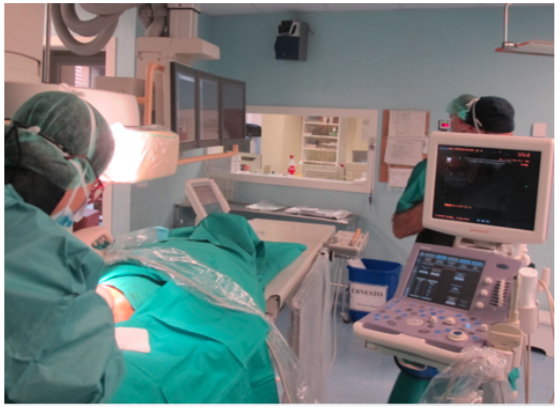
Explicar de una manera sencilla como se realiza una punción guiada por ecografía en la cateterización de la vena yugular interna utilizando la técnica de Seldinger.
Describir la metodología para la construcción de una fantoma vascular (cervical - yugulo/carotídeo) con la utilización de gelatina balística al 10%.
El objetivo principal es que radiólogos sin experiencia en procedimientos intervencionistas puedan conocer las técnicas utilizadas y adecuadas para evitar futuras complicaciones y poder prácticar con un fantoma que se asemeje a la anatomía vascular normal en la región cervical.
Descargas
Citas
Kim MC, Kim KS, Choi YK, et al. An estimation of right- and left-sided central venous catheter insertion depth using measurement of surface landmarks along the course of central veins. Anesth Analg. 2011 Jun;112(6):1371-4. doi: 10.1213/ANE.0b013e31820902bf. Epub 2011 Jan 13. PubMed PMID: 21233490.
Bannon MP, Heller SF, Rivera M. Anatomic considerations for central venous cannulation. Risk Manag Healthc Policy. 2011;4:27-39. doi: 0.2147/RMHP.S10383. Epub 2011 Apr 13. PubMed PMID: 22312225; PubMed Central PMCID: PMC3270925.
Brass P, Hellmich M, Kolodziej L, et al. Ultrasound guidance versus anatomical landmarks for internal jugular vein catheterization. Cochrane Database Syst Rev. 2015 Jan 9;1:CD006962. doi: 10.1002/14651858.CD006962.pub2. Review. PubMed PMID: 25575244.
McGee DC, Gould MK. Preventing complications of central venous catheterization. N Engl J Med. 2003 Mar 20;348(12):1123-33. Review. PubMed PMID: 12646670.
Serafimidis K, Sakorafas GH, Konstantoudakis G, et al. Ultrasound-guided catheterization of the internal jugular vein in oncologic patients; comparison with the classical anatomic landmark technique: a prospective study. Int J Surg. 2009 Dec;7(6):526-8. doi: 10.1016/j.ijsu.2009.08.011. Epub 2009 Sep 12. PubMed PMID: 19751852.
Nolting L, Hunt P, Cook T, Douglas B. An Inexpensive and Easy Ultrasound Phantom: A Novel Use for SPAM. J Ultrasound Med. 2016 Mar 3. pii: 14.06023. [Epub ahead of print] PubMed PMID: 26939600.
Schmidt GA, Maizel J, Slama M. Ultrasound-guided central venous access: what's new? Intensive Care Med. 2015 Apr;41(4):705-7. doi: 10.1007/s00134-014-3628-6. Epub 2015 Jan 8. Review. PubMed PMID: 25567384.
Hocking G, Hebard S, Mitchell CH. Review of the benefits and pitfalls of phantoms in ultrasound-guided regional anesthesia. Reg Anesth Pain Med 2011; 36:162–170.
Shiels WE. Soft tissue foreign bodies: sonographic diagnosis and therapeutic management. Ultrasound Clin 2007; 2:669–681.
American College of Emergency Physicians. Emergency ultrasound guidelines. Ann Emerg Med 2009; 53:550–570.