Valoración de las técnicas de imagen en la determinación de la penumbra isquémica en pacientes con ictus isquémico agudo
Palabras clave:
penumbra isquémica, poster, seram, ictus isquémico agudoResumen
Objetivos Docentes
• Definir el concepto de penumbra isquémica.
• Describir las distintas modalidades propuestas para determinar el parénquima infartado y la penumbra isquémica basadas en técnicas de imagen.
• Discutir las posibles ventajas y limitaciones de cada una de ellas.
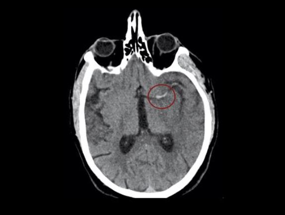
• Presentar ejemplos de casos con las técnicas descritas.
Revisión del tema
Introducción.
En el contexto del ictus agudo, el tiempo desde el inicio de los síntomas hasta el momento de la recanalización del vaso representa uno de los principales factores pronósticos (1).
Para el tratamiento con trombolisis endovenosa, la ventana terapéutica se basa en el tiempo de evolución (con el límite de 4,5 horas)(2).
Descargas
Citas
Lees KR, luhmki E, von Kummer R, Brott TG, Toni D, Grotta JC, et al;ECASS, ATLANTIS, NINDS and EPITHET rt-PA Study Group. Time to treatment with intravenous alteplase and outcome in stroke: an updated pooled analysis of ECASS, ATLANTIS, NINDS, and EPITHET trials. Lancet. 2010;375:1695–1703.
2015 AHA/ASA Focused Update of the 2013 Guidelines for the Early Management of Patients With Acute Ischemic Stroke Regarding Endovascular Treatment A Guideline for Healthcare Professionals From the American Heart Association/American Stroke Association. Stroke. 2015;46:000-000.
Jovin TG, Chamorro A, Davalos A. et al . Thrombectomy within 8 hours after symptom onset in ischemic stroke. N Engl J Med. 2015;372:2296-2306.
Ebinger M, De Silva DA, Christensen S, Parsons MW, Markus R, Donnan GA, Davis SM. J Clin Neurosci. 2009 Feb;16(2):178-87.
Rosso, C., & Samson, Y. (2014). The ischemic penumbra: the location rather than the volume of recovery determines outcome. Current opinion in neurology,27(1), 35-41.
Leiva-Salinas C, Wintermark M. Imaging of Ischemic Stroke. Neuroimaging clinics of North America. 2010;20(4):455-468. doi:10.1016/j.nic.2010.07.002.
Birenbaum D, Bancroft LW, Felsberg GJ. Imaging in Acute Stroke. Western Journal of Emergency Medicine. 2011;12(1):67-76.
Sobesky J. Refining the mismatch concept in acute stroke: lessons learned from PET and MRI. Journal of Cerebral Blood Flow & Metabolism. 2012;32(7):1416-1425.
Hirano, T. Searching for Salvageable Brain: The Detection of Ischemic Penumbra Using Various Imaging Modalities? Journal of Stroke and Cerebrovascular Diseases. 2014; 23(5), 795–798.
Best, A. C., Acosta, N. R., Fraser, J. E., Borges, M. T., Brega, K. E., Anderson, T., ... & Bert, R. J. (2012). Recognizing false ischemic penumbras in CT brain perfusion studies. Radiographics, 32(4), 1179-1196.
Wintermark, M., Flanders, A. E., Velthuis, B., Meuli, R., Van Leeuwen, M., Goldsher, D., ... & Anderson, J. (2006). Perfusion-CT assessment of infarct core and penumbra receiver operating characteristic curve analysis in 130 patients suspected of acute hemispheric stroke. Stroke, 37(4), 979-985.
Tan, J. C., Dillon, W. P., Liu, S., Adler, F., Smith, W. S., & Wintermark, M. (2007). Systematic comparison of perfusion-CT and CT-angiography in acute stroke patients. Annals of neurology, 61(6), 533-543.
Menon BK, Goyal M. Imaging Paradigms in Acute Ischemic Stroke: A Pragmtic Evidence-based Approach. Radiology. 2015 Oct;277(1):7-12
Donnan G, Baron JC, Davis S. The Ischemic Penumbra. CRC Press; 2007. p 246.
Kamalian, S., Kamalian, S., Boulter, D. J., Lev, M. H., Gonzalez, R. G., & Schaefer, P. W. Stroke differential diagnosis and mimics: Part 1. Applied Radiology. Nov 2015.
Srinivasan, A., Goyal, M., Azri, F. A., & Lum, C. State-of-the-art imaging of acute stroke. Radiographics. 2006; 26, S75-S95.
Mair G, Wardlaw JM. Imaging of acute stroke prior to treatment: current practice and evolving techniques. The British Journal of Radiology. 2014;87(1040):20140216.
Chalela JA, Kidwell CS, Nentwich LM, Luby M, Butman JA, Demchuk AM, Hill MD, Patronas N, Latour L, Warach S. Magnetic resonance imaging and computed tomography in emergency assessment of patients with suspected acute stroke: a prospective comparison. Lancet. 2007 Jan 27;369(9558):293-8.
Lansberg MG, Thijs VN, Hamilton S, Schlaug G, Bammer R, Kemp S, Albers GW; DEFUSE Investigators.. Evaluation of the clinical-diffusion and perfusion-diffusion mismatch models in DEFUSE. Stroke. 2007 Jun;38(6):1826-30.
Barber PA, Davis SM, Darby DG, Desmond PM, Gerraty RP, Yang Q, Jolley D, Donnan GA, Tress BM Absent middle cerebral artery flow predicts the presence and evolution of the ischemic penumbra. Neurology. 1999 Apr 12;52(6):1125-32.
Mishra NK, Albers GW, Christensen S, Marks M, Hamilton S, Straka M, Liggins JT, Kemp S, Mlynash M, Bammer R, Lansberg MG; Diffusion and Perfusion Imaging Evaluation for Understanding Stroke Evolution 2 Investigators. Comparison of magnetic resonance imaging mismatch criteria to select patients for endovascular stroke therapy. Stroke. 2014 May;45(5):1369-74.
Song SS, Latour LL, Ritter CH, Wu O, Tighiouart M, Hernandez DA, Ku KD, Luby M, Warach S. A pragmatic approach using magnetic resonance imaging to treat ischemic strokes of unknown onset time in a thrombolytic trial. Stroke. 2012 Sep;43(9):2331-5.
Geraldo AF, Berner LP, Haesebaert J, Chabrol A, Cho TH, Derex L, Hermier M, Louis-Tisserand G, Chamard L, Klaerke Mikkelsen I, Ribe L, Østergaard L, Hjort N, Pedraza S, Thomalla G, Baron JC, Nighoghossian N, Berthèzene Y. Does b1000-b0 Mismatch Challenge Diffusion-Weighted Imaging-Fluid Attenuated Inversion Recovery Mismatch in Stroke?. Stroke. 2016 Mar;47(3):877-81.
Lou M, Chen Z, Wan J, Hu H, Cai X, Shi Z, Sun J Susceptibility-diffusion mismatch predicts thrombolytic outcomes: a retrospective cohort study. AJNR Am J Neuroradiol. 2014 Nov-Dec;35(11):2061-7.