Patología del Espacio Supraclavicular en la edad pediátrica.
Palabras clave:
Patología del Espacio Supraclavicular, HSCResumen
Objetivos Docentes
Describir la anatomía del espacio supraclavicular, siendo éste un lugar de cruce entre numerosas estructuras.
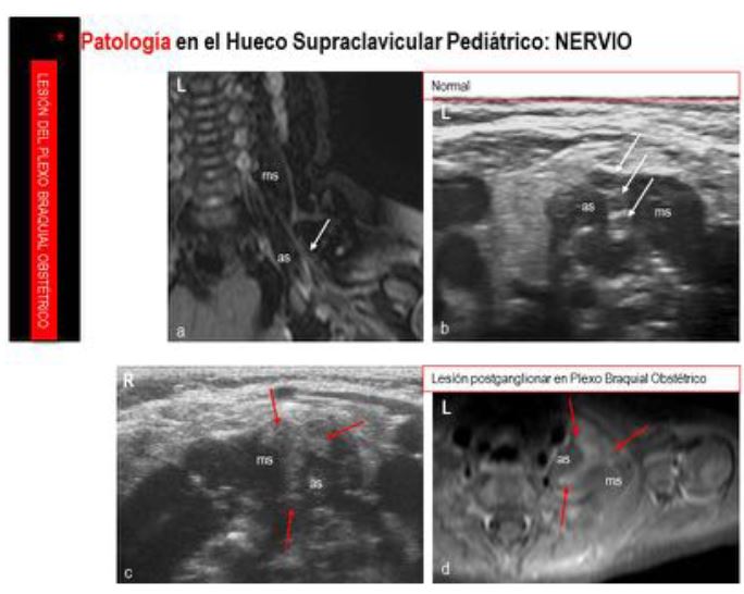
Correlacionar los elementos anatómicos del hueco supraclavicular (HSC) con la patología que en él se puede presentar.
Revisión del tema
Revisión del tema
Se seguirá el siguiente esquema (Fig.1):
• ANATOMÍA. Descripción anatómica del HSC.
• PATOLOGÍA. Galería de patología en este espacio.
• ANATOMÍA. Descripción anatómica del HSC (Fig.2)
Descargas
Citas
-Kumar R. Lindell M, Madewell J, David R, Swischuk LE. The clavicle: Normal and abnormal. RadioGraphics1989;9: 677-706.
-Meuwly JY, Lepori D, Theumann N, Schnyder P, Etechami G, Hohlfeld J, Gudinchet F. Multimodality Imaging Evaluation of the Pediatric Neck: Techniques and Spectrum of Findings. Radiographics 2005;25:931-48.
-Daniela Binaghi. Image Studies : When They can Help?. In Treatment of Peripheral Nerve Lesions. Basic Principles for General Neurosurgeons. Ed. Siqueira, Socolovsky, Malessy, Devi. PRISM BOOKS PVT LTD, 2011.
-ISSVA classification of vascular anomalies, except from Color Atlas of Vascular Tumors and Vascular Malformations, Cambridge University Press 978-0-521-84851-0 - Color Atlas of Vascular Tumors and Vascular Malformations Odile Enjolras, Michel Wassef and Rene Chapot -Koeller K, Alamo L. Congenital cystic masses of the neck: Radio-Pathologic correlation. Radiographics 1999;19:121-46.
-Laffan E, Ngan BY, Navarro OM. Pediatric Soft-Tissue Tumors and Pseudotumors: MR Imaging Features with Pathologic Correlation Part 2. Tumors of Fibroblastic/Myofibroblastic, So-called Fibrohistiocytic, Muscular, Lymphomatous, Neurogenic, Hair Matrix, and Uncertain Origin. RadioGraphics 2009; e36 • Published online 10.1148/rg.e36.
-Georgalas Ch, Kapoor , Chau H, Bhattacharyya A. Inflammatory focal myositis of the sternomastoid muscle: is there an absolute indication for biopsy? A case report and review of the literature. Eur Arch Otorhinolaryngol 2006;2:149-51.
-Nozaki T, Nosaka S, Miyazaki O, et al. Syndromes Associated with Vascular Tumors and Malformations: A Pictorial Review. RadioGraphics 2013;33:175-95.
-Martínez León MI. Chronic Recurrent Multifocal Osteomyelitis. In Learning Pediatric Diagnostic Imaging. Ed. Springer. 2011.
-Martínez León MI, Weil Lara B, Herrero Hernández A, Ceres Ruiz L. El síndrome del hueso evanescente. Radiología 2001;43:439-44.
-Mar WA, Taljanovic MS, Bagatell R, Graham AR, Speer DP, Hunter TB et al. Update on Imaging and Treatment of Ewing´s sarcoma Family Tumors: What the radiologist needs to know?. J Comput Assist Tomogr 2008;32:108-18.
-Lonergan GL, Schwab CM, Suarez ES, Carlson CL. Neuroblastoma, Ganglioneuroblastoma, and Ganglioneuroma: RadiologicPathologic Correlation. RadioGraphics 2002;22:911-34.
-Zhang WD, Chen YF, Li CX, Zhang L, Xu ZB, Zhang FJ. Computed tomography and magnetic resonance imaging findings of peripheral primitive neuroectodermal tumors of the head and neck. Eur J Radiol 2011;80:607-11.