MALFORMACIONES CONGÉNITAS DE LOS VASOS PULMONARES EN EL ADULTO
Palabras clave:
MALFORMACIONES CONGÉNITAS DE LOS VASOS PULMONARES, poster, seramResumen
Objetivos Docentes
• Conocer las manifestaciones radiológicas de estas malformaciones.
• Conocer las técnicas de imagen y reconstrucciones óptimas para su valoración.
• Conocer el impacto clínico y posible tratamiento de estas anomalías.
Revisión del tema
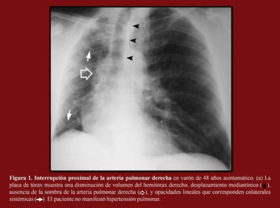
Las malformaciones congénitas pulmonares son raras y pueden clasificarse en tres categorías: anomalías broncopulmonares, anomalías vasculares aisladas y anomalías combinadas vasculares y pulmonares. Generalmente se detectan en el periodo prenatal/neonatal o primera infancia; sin embargo algunas permanecen asintomáticas y son detectadas incidentalmente en la edad adulta.
Estas malformaciones tienen manifestaciones radiológicas características, aunque pueden simular otras patologías y son causa frecuente de error diagnóstico. La caracterización de las lesiones congénitas normalmente se realiza mediante tomografía computarizada (TCMD) o resonancia magnética (RM) con contraste endovenoso y reconstrucciones 3D y en diferentes planos.
Descargas
Citas
Konen E, Raviv-Zilka L, Cohen RA, et al. Congenital pulmonary venolobar syndrome: spectrum of helical CT findings with emphasis on computerized reformatting. Radiographics. 2003 Sep-Oct;23(5):1175-84.
Castañer E, Gallardo X, Rimola J, et al. Congenital and acquired pulmonary artery anomalies in the adult: radiologic overview. Radiographics. 2006 Mar-Apr;26(2):349-71.
Thacker PG, Rao AG, Hill JG, Lee EY. Congenital lung anomalies in children and adults: current concepts and imaging findings. Radiol Clin North Am. 2014 Jan;52(1):155-81. doi: 10.1016/j.rcl.2013.09.001. Epub 2013 Oct 2.
White CS, Baffa JM, Haney PJ, Pace ME, Campbell AB. MR imaging of congenital anomalies of the thoracic veins. Radiographics. 1997 May-Jun;17(3):595-608.
Trotman-Dickenson B. Congenital lung disease in the adult: guide to the evaluation and management. J Thorac Imaging. 2015 Jan;30(1):46-59. doi: 10.1097/RTI.0000000000000127.
Brett W. Carter 1, John P. LichtenbergerIII 2 and Carol C. Congenital Abnormalities of the Pulmonary Arteries in Adults. AJR April 2014, Volume 202, Number 4
4. Morgan PW, Foley DW, Erickson SJ. Proximal interruption of a main pulmonary artery with transpleural collateral vessels: CT and MR appear- ances. J Comput Assist Tomogr 1991;15:311– 313.
Legras A, Guinet C, Alifano M, Lepilliez A, Régnard JF. A case of variant scimitar syndrome. Chest. 2012 Oct;142(4):1039-41. doi: 10.1378/chest.11-2732.
Lee EY, Dillon JE, Callahan MJ, et al. 3D multide- tector CT angiographic evaluation of extralobar pulmonary sequestration with anomalous venous drainage into the left internal mammary vein in a paediatric patient. Br J Radiol 2006;79(945): e99–102.
Lee EY, Tracy DA, Mahmood SA, et al. Preoperative MDCT evaluation of congenital lung anomalies in children: comparison of axial, multiplanar, and 3D images. AJR Am J Roentgenol 2011;196(5): 1040–6.
Kang M, Khandelwal N, Ojili V, et al. Multidetector CT angiography in pulmonary sequestration. J Comput Assist Tomogr 2006;30(6):926–32.
Zylak CJ, Eyler WR, Spizarny DL, Stone CH. Developmental lung anomalies in the adult: radiologic-pathologic correlation. RadioGraphics 2002; 22(spec no):S25–S43.


