Correlación del grado de respuesta tras quimioterapia neoadyuvante, en pacientes con Ca de mama con fenotipo triple negativo. RM vs anatomía patológica.
Palabras clave:
grado de respuesta, quimioterapia, neoadyuvante, fenotipo triple negativo, cáncer de mama, poster, seramResumen
Objetivos
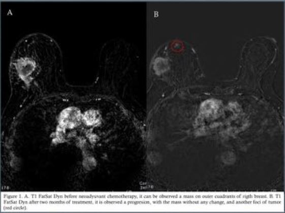
La quimioterapia sistémica esta ampliamente aceptada y usada en el tratamiento del cáncer de mama, mas aun en aquellas pacientes con cáncer localmente avanzado(1-4). Últimamente se ha demostrado que la quimioterapia neoadyuvante en estos pacientes con tumoración localmente avanzada, tienen tiempo libre de enfermedad y supervivencia similar a aquellas pacientes tratadas únicamente con quimioterapia convencional(5), pero si se ha demostrado que estas paciente son subsidiarias de cirugía conservadora de la mama(6-8).
La RM, con secuencias anatómicas, funcionales y tras al administración de contraste intravenoso, es ampliamente superior que la mamografía y la ultrasonografía en el estudio de la tumoración en cuanto a dimensiones, características y multifocalidad(4,9-11). También se ha demostrado que la RM previo y
posterior al tratamiento es un marcador mas seguro que la exploración física, para determinar la repuesta tumoral(4,12,13).
La repuesta del cáncer de mama a la neoadyuvancia viene determinada por muchos factores, entre ellos la inmunohistoquímica específica de cada tumor(luminal A, luminal B, HER2+, triple negativo), evidenciándose mejor repuesta en aquellas pacientes con fenotipo triple negativo o HER2+no
luminal(14-16).
Descargas
Citas
Marinovich ML, Sardanelli F, Ciatto S, Mamounas E, Brennan M, Macaskill P, et al. Early prediction of pathologic response to neoadjuvant therapy in breast cancer: Systematic review of the accuracy of MRI. Breast [Internet]. Elsevier Ltd; 2012;21(5):669–77. Available from: http://dx.doi.org/10.1016/j.breast.2012.07.006
Jung H, Baek H, Ahn J, Baek S. Prediction of pathologic response to neoadjuvant chemotherapy in patients with breast cancer using diffusion-weighted imaging and MRS. NMR Biomed. 2012;25:1349–59.
Ojeda-Fournier H, de Guzman J, Hylton N. Breast Magnetic Resonance Imaging for Monitoring Response to Therapy. Magn Reson Imaging Clin N Am [Internet]. Elsevier Inc; 2013;21(3):533–46. Available from: http://dx.doi.org/10.1016/j.mric.2013.04.005
Hylton NM, Blume JD, Bernreuter WK, Pisano ED, Rosen MA, Morris EA, et al. Locally Advanced Breast Cancer: MR Imaging for Prediction of Response to Neoadjuvant Chemotherapy--Results from ACRIN 6657/I-SPY TRIAL. Radiology. 2012;263(3):663–72.
Lobbes MBI, Prevos R, Smidt M, Tjan-Heijnen VCG, van Goethem M, Schipper R, et al. The role of magnetic resonance imaging in assessing residual disease and pathologic complete response in breast cancer patients receiving neoadjuvant chemotherapy: a systematic review. Insights Imaging [Internet]. 2013;4(2):163–75. Available from: http://www.pubmedcentral.nih.gov/articlerender.fcgi?artid=3609956&tool=pmcentrez&rendertype=abstract
Marinovich ML, Houssami N, MacAskill P, Sardanelli F, Irwig L, Mamounas EP, et al. Meta-analysis of magnetic resonance imaging in detecting residual breast cancer after neoadjuvant therapy. J Natl Cancer Inst. 2013;105(February 2011):321–33.
Marcos de Paz LM, Tejerina Bernal A, Arranz Merino ML, Calvo de Juan V. Breast MR imaging changes after neoadjuvant chemotherapy: correlation with molecular subtypes. Radiologia [Internet]. 2012;54(5):442–8. Available from: http://www.ncbi.nlm.nih.gov/pubmed/21937065
Chen JH, Carpenter PM, Mehta RS, Nalcioglu O. MRI Evaluation of Pathologically Complete Response and Residual Tumors in Breast Cancer After Neoadjuvant Chemotherapy. Am Cancer Soc. 2008;112(November 2007):17–26.
Wu L-M, Hu J-N, Gu H-Y, Hua J, Chen J, Xu J-R. Can diffusion-weighted MR imaging and contrast-enhanced MR imaging precisely evaluate and predict pathological response to neoadjuvant chemotherapy in patients with breast cancer? Breast Cancer Res Treat. 2012;135(1):17–28.
Richard R, Thomassin I, Chapellier M, Scemama A, Bazelaire C De. Diffusion-weighted MRI in pretreatment prediction of response to neoadjuvant chemotherapy in patients with breast cancer. Eur Radiol. 2013;23:2420–31.
Partridge SC, McDonald ES. Diffusion Weighted Magnetic Resonance Imaging of the Breast: Protocol Optimization, Interpretation, and Clinical Applications. Magn Reson Imaging Clin N Am [Internet]. Elsevier Inc; 2013;21(3):601–24. Available from: http://dx.doi.org/10.1016/j.mric.2013.04.007
Williams M, Eatrides J, Kim J, Talwar H, Esposito N, Szabunio M, et al. Comparison of breast magnetic resonance imaging clinical tumor size with pathologic tumor size in patients status post-neoadjuvant chemotherapy. Am J Surg [Internet]. Elsevier Inc; 2013;206(4):567–73. Available from: http://dx.doi.org/10.1016/j.amjsurg.2013.02.006
Bolan PJ. Magnetic Resonance Spectroscopy of the Breast Current Status. Magn Reson Imaging Clin NA [Internet]. Elsevier Inc; 2013;21(3):625–39. Available from: http://dx.doi.org/10.1016/j.mric.2013.04.008
Ko ES, Han B, Kim RB, Ko EY, Shin JH, Hahn SY, et al. Analysis of factors that influence the accuracy of magnetic resonance imaging for predicting response after neoadjuvant chemotherapy in locally advanced breast cancer. Ann Surg Oncol [Internet]. 2013;20(8):2562–8. Available from: http://www.ncbi.nlm.nih.gov/pubmed/23463090
Lips EH, Mukhtar R a., Yau C, De Ronde JJ, Livasy C, Carey LA, et al. Lobular histology and response to neoadjuvant chemotherapy in invasive breast cancer. Breast Cancer Res Treat. 2012;136(1):35–43.
McGuire KP, Toro-Burguete J, Dang H, Young J, Soran A, Zuley M, et al. MRI staging after neoadjuvant chemotherapy for breast cancer: does tumor biology affect accuracy? Ann Surg Oncol [Internet]. 2011;18(11):3149–54. Available from: http://www.ncbi.nlm.nih.gov/pubmed/21947592
Rigter LS, Loo CE, Linn SC, Sonke GS, Werkhoven E Van, Lips EH, et al. Neoadjuvant chemotherapy adaptation and serial MRI response monitoring in ER-positive HER2-negative breast cancer. Br J Cancer [Internet]. Nature Publishing Group; 2013;Dec 10(109(12)):2965–72. Available from: http://dx.doi.org/10.1038/bjc.2013.661
Wilmes LJ, Mclaughlin RL, Newitt DC, Singer L, Sinha SP, Proctor E, et al. High-Resolution Diffusion-Wighted Imaging for Monitoring Breast Cancer Treatment Response. Acad Radiol [Internet]. Elsevier Ltd; 2013;20(5):581–9. Available from: http://dx.doi.org/10.1016/j.acra.2013.01.009
P Mukherjee, S Sharma, ZA Sheikh, DK Vijaykumar Correlation of clinico-pathologic and radiologic parameters of response to neoadjuvant chemotherapy in breast cancer Indian Journal of Cancer, Vol. 51, No. 1, January-March, 2014, pp. 25-29
Manuscript A. NIH Public Access. Changes. 2012;29(6):997–1003.


