VALORACIÓN DE LA COLUMNA TORACOLUMBAR POSTOPERADA Y SUS COMPLICACIONES
Palabras clave:
columna, toracolumbar, toraxlumbar, complicaciones, vertebralResumen
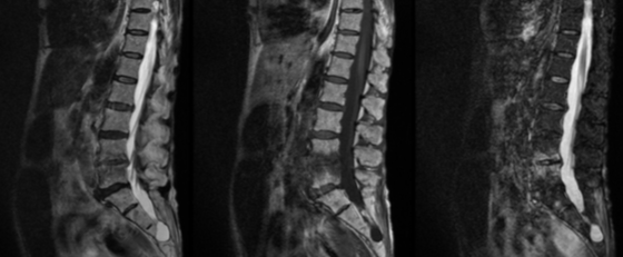
Hemos estudiado una serie de 40 pacientes que necesitaron intervención de la columna toracolumbar. A través del análisis de esta serie vamos a describir y mostrar los hallazgos en imagen de la columna postoperada normal y las características en imagen del material y dispositivos quirúrgicos. Como segundo objetivo nos hemos propuesto ilustrar las diferentes complicaciones de la cirugía realizando un enfoque multimodal con radiología simple, Tomografía Computarizada Multicorte y Resonancia Magnética de 1.5 y 3T.
Material y métodos
Se trata de un estudio retrospectivo y descriptivo. Se revisaron las Resonancias Magnéticas realizadas en el periodo 2014-2015 a pacientes que sufrieron una intervención en la columna toracolumbar. Se seleccionaron 40 casos, de los cuales 29 presentaban algún tipo de complicación (72,5%). Se incluyen aquellos pacientes que disponen de estudios de imagen tanto previos a la cirugía como posteriores. Entre los estudios de imagen evaluados están radiografías, tomografías computerizadas y resonancias magnéticas con equipos de 1,5 y 3T, tanto previos como posteriores a la cirugía.
Descargas
Citas
Complications of Spinal Instrumentation. Radiographics 2006. Phillip M. Young, MD, Thomas H. Berquist, MD, Laura W. Bancroft, MD, and Jeffrey J. Peterson, MD.
Lumbar Spine Fusion and Stabilization: Hardware, Techniques, and Imaging Appearances. Radiographics 2007. Elizabeth E. Rutherford, FRCR, Linda J. Tarplett,
RGN, ONC, Evan M. Davies, FRCS, John M. Harley, FRCS, and Leonard J. King, FRCR.
Normal and Abnormal Imaging Findings in Lumbar Total Disk Replacement: Devices and Complications. Radiographics 2008. Ryan D. Murtagh, MD, MBA, Robert M.
Quencer, MD, Dan S. Cohen, MD, James J. Yue, MD, and Evelyn L. Sklar, MD.
Imaging Features of Postoperative Complications After Spinal Surgery and Instrumentation. AJR 2012. Daichi Hayashi, Frank W. Roemer, Asim Mian, Monther Gharaibeh, Bernhard Müller, and Ali Guermazi.
Imaging of Current Spinal Hardware: Lumbar Spine. AJR 2014. Alice S. Ha1 and Jonelle M. Petscavage-Thomas.
Spinal fixation. Part 3. Complications of spinal instrumentation. Radiographics 1993. Slone RM, MacMillan M, Montgomery WJ.
Spinal fixation. Part 1. Principles, basic hardware, and fixation techniques for the cervical spine. Radiographics 1993. Slone RM, MacMillan M, Montgomery WJ.
Spinal fixation. Part 2. Fixation techniques and hardware for the thoracic and lumbosacral spine. Radiographics 1993. Slone RM1, MacMillan M, Montgomery WJ, Heare M.
Orthopedic fixation devices. Radiographics 1991. Slone RM, Heare MM, Vander Griend RA, Montgomery WJ.
Principles and imaging of spinal instrumentation. Radiographics 1995. Slone RM, McEnery KW, Bridwell KH, Montgomery WJ.
Fixation techniques and instrumentation used in the thoracic, lumbar, and lumbosacral spine. Radiographics 1995. Slone RM, McEnery KW, Bridwell KH, Montgomery WJ.
Rutherford EE, Tarplett LJ, Davies EM, Harley JM, King LJ. Lumbar spine fusion and stabilization: hardware, techniques, and imaging appearances. RadioGraphics 2007.