PETTC y PETRM en el manejo de los tumores de cabeza y cuello
Palabras clave:
PET, TC, RM, tumores cabeza, cuello, diagn´sticoResumen
Describir las situaciones clínicas en donde las técnicas de imagen morfológicas y metabólicas pueden contribuir a un mejor manejo de los tumores de cabeza y cuello
Explicar el rendimiento de la PETTC y PETRM en estas situaciones clínicas, tanto de forma individual como conjunta.
Revisión del tema
Los tumores de cabeza y cuello suponen en torno a un 5% del total de los cánceres diagnosticados. España presenta una elevada incidencia de cánceres de vías aerodigestivas superiores (CVADS). Cabeza y cuello es una región anatómica compleja, los tumores pueden asentar en múltiples localizaciones y, por tanto, presentar una clínica muy heterogénea. Todo esto implica una serie de dificultades en el proceso diagnóstico, lo que va a repercutir negativamente en el pronóstico de los pacientes.
El diagnóstico de estas neoplasias se basa fundamentalmente en:
La valoración de la sintomatología.
La exploración física, ya que la mayoría de estos tumores asientan en las mucosas.
Pruebas de imagen complementarias
La confirmación histológica o citológica, que es el diagnóstico de certeza de estos tumores.
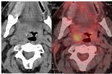
Aunque tradicionalmente las técnicas de imagen morfológicas como la TC, la RM y la ecografía han resultado de gran utilidad a los clínicos para el manejo de la enfermedad oncológica, éstos han encontrado en la PET una información metabólica adicional. Considerando estas diferencias, la PET
resulta de mayor utilidad para la distinguir tumores malignos de procesos benignos, para determinar el grado de malignidad de las lesiones, para monitorizar la respuesta al tratamiento y planificar los volúmenes de radioterapia.
Las indicaciones de la PETTAC en tumores de cabeza y cuello son las siguientes:
A) Estadificación
B) Tumor de origen desconocido
C) Valoración de la respuesta a los tratamientos
D) Detección de recidivas
Descargas
Citas
SUVmax on FDGPET is a predictor of prognosis in patients with maxillary sinus cancer.
Doi H, Kitajima K, Fukushima K, Kawanaka Y, Mouri M, Yamamoto S, Ishikura R, Terada T, Noguchi K, Hirota S.Jpn J Radiol. 2016 Feb 2
Dynamic contrastenhanced MRI, diffusionweighted MRI and 18FFDG PET/CT for the prediction of survival in oropharyngeal or hypopharyngeal squamous cell carcinoma treated with chemoradiation.
Ng SH, Liao CT, Lin CY, Chan SC, Lin YC, Yen TC, Chang JT, Ko SF, Fan KH, Wang HM, Yang LY, Wang JJ. Eur Radiol. 2016 Feb 24
18FFDG PETCT performed before and during radiation therapy of head and neck squamous cell carcinoma: Are they independent or complementary to each other?
Min M, Lin P, Lee M, Shon IH, Lin M, Forstner D, Tieu MT, Chicco A, Bray V, Fowler A. J Med Imaging Radiat Oncol. 2016 Feb 12
Prognostic Value of 2[18F] Fluoro2deoxyDglucose Positron Emission TomographyComputed Tomography Scan Carried out During and After Radiation Therapy for Head and Neck Cancer Using Visual Therapy Response Interpretation Criteria.
Min M, Lin P, Lee M, Ho Shon I, Lin M, Forstner D, Tieu MT, Chicco A, Bray V, Fowler A. Clin Oncol (R Coll Radiol). 2016 Jan 9
Combined multimodal coregistration of PET/CT and MRI images increases diagnostic accuracy in squamous cell carcinoma staging.
Stecco A, Ciolfi S, Buemi F, Cassarà A, Sacchetti GM, Brambilla M, Carriero A. Radiol Med. 2016 Jan 11.
Detecting Residual/Recurrent Head Neck Squamous Cell Carcinomas Using PET or PET/CT: Systematic Review and Metaanalysis.
Cheung PK, Chin RY, Eslick GD. Otolaryngol Head Neck Surg. 2016 Mar;154(3):42132
Combined PET/CTperfusion in patients with head and neck cancers might predict failure after radiochemotherapy: a proof of concept study.
Pietsch C, de Galiza Barbosa F, Hüllner MW, Schmid DT, Haerle SK, Huber GF, Studer G, Hany TF, VeitHaibach P. BMC Med Imaging. 2015 Dec 29;15:60
Predicting extracapsular spread of head and neck cancers using different imaging techniques: a systematic review and metaanalysis.
Su Z, Duan Z, Pan W, Wu C, Jia Y, Han B, Li C. Int J Oral Maxillofac Surg. 2015 Dec 17. pii: S09015027(15)014344
Imaging strategy for response evaluation to chemoradiotherapy of the nodal disease in patients with head and neck squamous cell carcinoma.
Nishimura G, Yabuki K, Hata M, Komatsu M, Taguchi T, Takahashi M, Shiono O, Sano D, Arai Y, Takahashi H, Chiba Y, Oridate N. Int J Clin Oncol. 2015 Dec 28.
18FFDG PET/CT to assess response and guide riskstratified followup after chemoradiotherapy for oropharyngeal squamous cell carcinoma.
Bird T, Barrington S, Thavaraj S, Jeannon JP, Lyons A, Oakley R, Simo R, Lei M, Guerrero Urbano T. Eur J Nucl Med Mol Imaging. 2015 Dec 28
The Clinical Usefulness of 18Ffluorodeoxyglucose Positron Emission Tomography to Predict Oncologic Outcomes and PETbased Radiotherapeutic Considerations in Locally Advanced Nasopharyngeal Carcinoma.
Yoon HI, Kim KH, Lee J, Roh YH, Yun M, Cho BC, Lee CG, Keum KC. Cancer Res Treat. 2015 Dec 11
[18F]FDG PET/CT imaging for detection of nodal metastases in patients with squamous cell carcinoma of the pharynx and larynx: comparison with CT.
Suenaga Y, Kitajima K, Kanda T, Otsuki N, Nibu KI, Sasaki R, Itoh T, Sugimura K. Jpn J Radiol. 2015 Dec 15
Use of 18FFludeoxyglucosePositron Emission Tomography/Computed Tomography for Patient Management and Outcome in Oropharyngeal Squamous Cell Carcinoma: A Review.
Taghipour M, Sheikhbahaei S, Marashdeh W, Solnes L, Kiess A, Subramaniam RM. JAMA Otolaryngol Head Neck Surg. 2016 Jan 1;142(1):7985
DiffusionWeighted MRI in the Assessment of Early Treatment Response in Patients with SquamousCell Carcinoma of the Head and Neck: Comparison with Morphological and PET/CT Findings.
Martins EB, Chojniak R, Kowalski LP, Nicolau UR, Lima EN, Bitencourt AG. PLoS One. 2015 Nov 12;10(11)
Comparison of (18)FFDG PET/CT, MRI and SPECT in the diagnosis of local residual/recurrent nasopharyngeal carcinoma: A metaanalysis.
Wei J, Pei S, Zhu X. Oral Oncol. 2016 Jan;52:117
18FFDG PET/CT for the diagnosis of residual or recurrent nasopharyngeal carcinoma after radiotherapy: a metaanalysis.
Zhou H, Shen G, Zhang W, Cai H, Zhou Y, Li L. J Nucl Med. 2015 Nov 5
Present and future role of FDGPET/CT imaging in the management of head and neck carcinoma. Kitajima K, Suenaga Y, Sugimura K. Jpn J Radiol. 2015 Dec;33(12):77689.
Characteristics and Limitations of FDG PET/CT for Imaging of Squamous Cell Carcinoma of the Head and Neck: A Comprehensive Review of Anatomy, Metastatic Pathways, and Image Findings.
Plaxton NA, Brandon DC, Corey AS, Harrison CE, Karagulle Kendi AT, Halkar RK, Barron BJ. AJR Am J Roentgenol. 2015 Nov;205(5):W51931
Intratherapy or Posttherapy FDG PET or FDG PET/CT for Patients With Head and Neck Cancer: A Systematic Review and Metaanalysis of Prognostic Studies.
Sheikhbahaei S, Ahn SJ, Moriarty E, Kang H, Fakhry C, Subramaniam RM. AJR Am J Roentgenol. 2015 Nov;205(5):110213
Effectiveness of an 18FFDGPET based strategy to optimize the diagnostic trajectory of suspected recurrent laryngeal carcinoma after radiotherapy: The RELAPS multicenter randomized trial.
de Bree R, van der Putten L, van Tinteren H, Wedman J, Oyen WJ, Janssen LM, van den Brekel MW, Comans EF, Pruim J, Takes RP, Hobbelink MG, Valdés Olmos R, van der Laan BF, Boers M, Hoekstra OS, Leemans CR. Radiother Oncol. 2015 Oct 15.
Screening for distant metastases in head and neck cancer patients using FDGPET and chest CT: validation of an algorithm.
Senft A, Hoekstra OS, Witte BI, Leemans CR, de Bree R. Eur Arch Otorhinolaryngol. 2015 Sep 9
Prognostic value of FDG PET/CT in head and neck squamous cell carcinomas.
Dequanter D, Shahla M, Aubert C, Deniz Y, Lothaire P. Onco Targets Ther. 2015 Aug 26;8:227983
Spatiotemporal stability of pretreatment 18FFludeoxyglucose uptake in head and neck squamous cell carcinomas sufficient for dose painting. Rasmussen JH, Vogelius IR, Aznar MC, Fischer BM, Christensen CB, Friborg J, Loft A, Kristensen CA, Bentzen SM, Specht L. Acta Oncol. 2015;54(9):141622
Postoperative PET/CT and target delineation before adjuvant radiotherapy in patients with oral cavity squamous cell carcinoma.
Dutta PR, Riaz N, McBride S, Morris LG, Patel S, Ganly I, Wong RJ, Palmer F, Schöder H, Lee N. Head Neck. 2015 Sep 3.