Diagnóstico radiológico de los tumores óseos primarios malignos de la columna vertebral
Palabras clave:
póster, seram, columna vertebral, tumor óseo primarioResumen
Objetivos
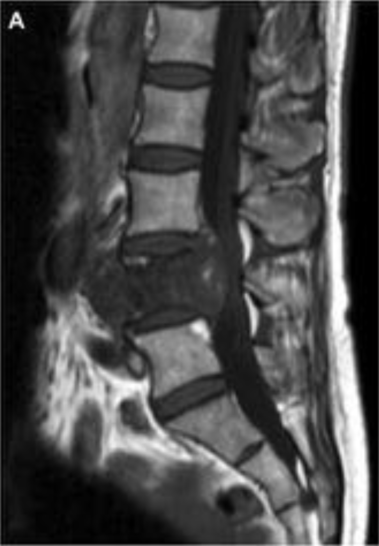
Revisar las características clínicas y radiológicas por TC y RM de los tumores óseos primarios de columna (TOPM), correlacionándolas con el diagnóstico de anatomía patológica definitivo.
Material y métodos
Se revisaron de forma retrospectiva los datos clínicos y radiológicos de 25 pacientes con diagnóstico histológico de TOPM de raquis visitados en nuestro hospital desde 1996 a 2015. Se excluyeron los casos de tumores de células gigantes, linfoma, y de enfermedad de células plasmáticas (plasmocitoma o mieloma).
Descargas
Citas
Aflatoon K, Staals E, Bertoni F, Bacchini P, Donati D, Fabbri N, et al. Hemangioendothelioma of the spine. Clin Orthop Relat Res. 2004 Jan;(418):191–7.
Farsad K, Kattapuram S V, Sacknoff R, Ono J, Nielsen GP. Sacral chordoma. Radiographics. Radiological Society of North America; 2009 Jan 1;29(5):1525–30.
Flemming DJ, Murphey MD. Enchondroma and Chondrosarcoma. Semin Musculoskelet Radiol; 2000 Mar;Volume 4(Number 1):0059–72.
Ilaslan H, Sundaram M, Unni KK, Dekutoski MB. Primary Ewing‘s sarcoma of the vertebral column. Skeletal Radiol. 2004;33:506–513.
Kahn SHM, De Schepper AM. Primary Tumors of the Osseous Spine. En: Van Goethem JWM, van den Hauwe L, Parizel PM, editores. Spinal Imaging. Diagnostic Imaging of the Spine and Spinal Cord. Germany: Springer; 2007. p.475-500.
Katonis P, Datsis G, Karantanas A, Kampouroglou A, Lianoudakis S, Licoudis S, et al. Spinal osteosarcoma. Clin Med Insights Oncol. 2013 Jan;7:199–208.
Larochelle O, Périgny M, Lagacé R, Dion N, Giguère C. Best cases from the AFIP: epithelioid hemangioendothelioma of bone. Radiographics. Jan;26(1):265–70.
Matamalas A, Gargallo A, Porcel JA, García de Frutos A, Pellisé F. Cervical spine epithelioid
hemangioendothelioma: case report. Eur Rev Med Pharmacol Sci. 2014 Jan;18(1 Suppl):72–5.
Murphey MD, Andrews CL, Flemming DJ, Temple HT, Smith WS, Smirniotopoulos JG. From the archives of the AFIP. Primary tumors of the spine: radiologic pathologic correlation. Radiographics. 1996 Sep 1;16(5):1131–58.
Murphey MD, Choi JJ, Kransdorf MJ, Flemming DJ, Gannon FH. Imaging of Osteochondroma: Variants and Complications with Radiologic-Pathologic Correlation1. RadioGraphics. Radiological Society of North America; 2000.
Rodallec MH, Feydy A, Larousserie F, Anract P, Campagna R, Babinet A, et al. Diagnostic imaging of solitary tumors of the spine: what to do and say. Radiographics. Radiological Society of North America; 2008 Jan;28(4):1019–41.
Smolders D, Wang X, Drevelengas A, Vanhoenacker F, De Schepper AM. Value of MRI in the diagnosis of non-clival, non-sacral chordoma. Skeletal Radiol. 2003; 32:343-350.
Van Goethem JW, van den Hauwe L, Ozsarlak O, De Schepper AM, Parizel PM. Spinal tumors. Eur J Radiol. 2004;50:159–176.
White LM, Kandel R. Osteoid-Producing Tumors of Bone. Semin Musculoskelet Radiol. 2000 Mar;Volume 4(Number 1):0025–44.
Wippold FJ, Koeller KK, Smirniotopoulos JG. Clinical and imaging features of cervical chordoma. AJR Am J Roentgenol. 1999;172:1423–1426.
York JE, Kaczaraj A, Abi-Said D, Fuller GN, Skibber JM, Janjan NA, Gokaslan ZL. Sacral chordoma: 40-year experience at a major cancer center. Neurosurgery. 1999;44:74–79.


