Precisión y eficiencia de un software automático para cuantificar los volúmenes del ventrículo izquierdo por cardio-resonancia magnética (CRM)
Palabras clave:
volúmenes del ventrículo izquierdo, volumen del ventrículo izquierdo, poster, seram, cardio-resonancia magnética, softwareResumen
Objetivos
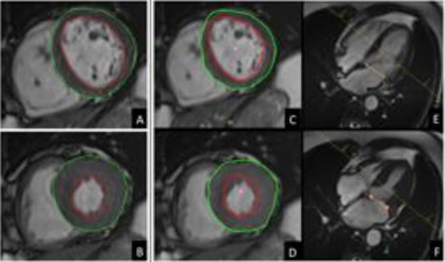
Evaluar la precisión, fiabilidad, y eficiencia temporal de un software de segmentación del ventrículo izquierdo totalmente automatizado recientemente comercializado, para calcular los volúmenes y función del VI, comparado con la segmentación manual convencional.
Material y métodos
Este estudio de cohortes consistió en 67 individuos consecutivos con enfermedad isquémica conocida o sospechada, a los que se les indicó una RM cardíaca de estrés con adenosina. Todos los pacientes estaban estables y en ritmo sinusal en el momento de la exploración.
Los pacientes con claustrofobia severa, objetos no compatibles con RM, portadores de marcapasos/DAI e insuficiencia renal (filtrado glomerular < 30 ml/min) fueron excluidos. A los pacientes se les indicó evitar alimentos y bebidas con cafeína en las 24h anteriores a la exploración. El comité institucional
aprobó el protocolo del estudio.
Descargas
Citas
Windecker S, Kolh P, Alfonso F, Collet JP, Cremer J, et al. 2014 ESC/EACTS Guidelines on myocardial revascularization: The Task Force on Myocardial Revascularization of the European Society of Cardiology (ESC) and the European Association for Cardio-Thoracic Surgery (EACTS)Developed with the special contribution of the European Association of Percutaneous Cardiovascular Interventions (EAPCI). European heart journal. 2014;35(37):2541-619.
Bruder O, Wagner A, Lombardi M, Schwitter J, van Rossum A, Pilz G, et al. European Cardiovascular Magnetic Resonance (EuroCMR) registry--multi national results from 57 centers in 15 countries. Journal of cardiovascular magnetic resonance : official journal of the Society for Cardiovascular Magnetic Resonance. 2013;15:9.
Kramer CM, Barkhausen J, Flamm SD, Kim RJ, Nagel E. Standardized cardiovascular magnetic resonance (CMR) protocols 2013 update. Journal of cardiovascular magnetic resonance : official journal of the Society for Cardiovascular Magnetic Resonance. 2013;15:91.
Hundley WG, Bluemke DA, Finn JP, Flamm SD, Fogel MA, Friedrich MG, et al. ACCF/ACR/AHA/NASCI/SCMR 2010 expert consensus document on cardiovascular magnetic resonance: a report of the American College of Cardiology Foundation Task Force on Expert Consensus Documents. Journal of the American College of Cardiology. 2010;55(23):2614-62.
Codella NC, Cham MD, Wong R, Chu C, Min JK, Prince MR, et al. Rapid and accurate left ventricular chamber quantification using a novel CMR segmentation algorithm: a clinical validation study. Journal of magnetic resonance imaging : JMRI. 2010;31(4):845-53.
Kawaji K, Codella NC, Prince MR, Chu CW, Shakoor A, LaBounty TM, et al. Automated segmentation of routine clinical cardiac magnetic resonance imaging for assessment of left ventricular diastolic dysfunction. Circulation Cardiovascular imaging. 2009;2(6):476-84.
Nassenstein K, de Greiff A, Hunold P. MR evaluation of left ventricular volumes and function: threshold-based 3D segmentation versus short-axis planimetry. Investigative radiology. 2009;44(10):635-40.
Francois CJ, Fieno DS, Shors SM, Finn JP. Left ventricular mass: manual and automatic segmentation of true FISP and FLASH cine MR images in dogs and pigs. Radiology. 2004;230(2):389-95.
Nguyen C, Kuoy E, Ruehm S, Krishnam M. Reliability and reproducibility of quantitative assessment of left ventricular function and volumes with 3-slice segmentation of cine steady-state free precession short axis images. European journal of radiology. 2015;84(7):1249-58.
Schulz-Menger J, Bluemke DA, Bremerich J, Flamm SD, Fogel MA, Friedrich MG, et al. Standardized image interpretation and post processing in cardiovascular magnetic resonance: Society for Cardiovascular Magnetic Resonance (SCMR) board of trustees task force on standardized post processing. Journal of cardiovascular magnetic resonance : official journal of the Society for Cardiovascular Magnetic Resonance. 2013;15:35.
Bland JM, Altman DG. Statistical methods for assessing agreement between two methods of clinical measurement. Lancet. 1986;1(8476):307-10.
Lin LI. A concordance correlation coefficient to evaluate reproducibility. Biometrics. 1989;45(1):255-68.
Bingham SE, Hachamovitch R. Incremental prognostic significance of combined cardiac magnetic resonance imaging, adenosine stress perfusion, delayed enhancement, and left ventricular function over preimaging information for the prediction of adverse events. Circulation. 2011;123(14):1509-18.
Kelly MJ, Thompson PL, Quinlan MF. Prognostic significance of left ventricular ejection fraction after acute myocardial infarction. A bedside radionuclide study. British heart journal. 1985;53(1):16-24.
Reinstadler SJ, Klug G, Feistritzer HJ, Kofler M, Pernter B, Gobel G, et al. Prognostic value of left ventricular global function index in patients after ST-segment elevation myocardial infarction. European heart journal cardiovascular Imaging. 2015.
Mazonakis M, Grinias E, Pagonidis K, Tziritas G, Damilakis J. Development and evaluation of a semiautomatic segmentation method for the estimation of LV parameters on cine MR images. Physics in medicine and biology. 2010;55(4):1127-40.
Papavassiliu T, Kuhl HP, Schroder M, Suselbeck T, Bondarenko O, Bohm CK, et al. Effect of endocardial trabeculae on left ventricular measurements and measurement reproducibility at cardiovascular MR imaging. Radiology. 2005;236(1):57-64.