LESIONES PSEUDOTUMORES DE TOBILLO Y PIE:
¿QUÉ DEBE SABER EL RADIÓLOGIO?
Palabras clave:
pseudotumores, tobillo, pie, lesiones pseudotumorales, poster, seramResumen
Objetivos Docentes
1.- Revisar las características en imagen de los pseudotumores del tobillo y pie.
2.- Exponer y describir los hallazgos radiológicos más representativos de las lesiones pseudotumorales de tobillo y pie diagnosticados en nuestro servicio.
Revisión del tema
Los tumores de tobillo y pie son frecuentes en la práctica clínica habitual. La gran mayoría de ellos son lesiones psedotumorales o tumores benignos.
Se revisan 100 casos de pseudotumores de tobillo y pie diagnosticados desde 2011.
Se exponen los casos más representativos de lesiones pseudotumorales encontrados en nuestro servicio:
músculos accesorios, gangliones/quistes sinoviales, neuroma de Morton, bursitis (intermetatarsiana, adventicia), lesiones cutáneas y subcutáneas (quiste epidermoide, granuloma anular), lesiones inflamatorias e infecciosas (nódulos reumatoides, tenosinovitis, abscesos), enfermedades metabólicas
(gota, calcicosis tumoral…) y otros pseudotumores (miositis osificante, fibromatosis plantar).
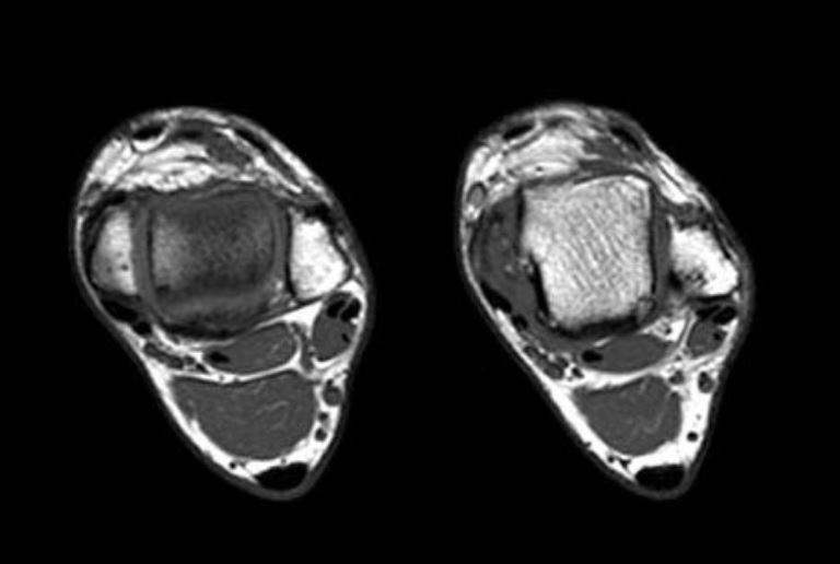
Se muestran imágenes ecográficas, primera prueba que se emplea para localizar la lesión y determinar su naturaleza, así como imágenes de RM que nos permite, filiar mejor la lesión, determinar la extensión y su relación con estructuras vecinas.
Descargas
Citas
Sookur PA, Naraghi AM, Bleakney RR, Jalan R, Chan O, White LM. Accessory muscles: anatomy, symptoms, and radiologic evaluation. Radiographics. 2008;28:481–499.
Kinoshita M, Okuda R, Morikawa J, Abe M. Tarsal tunnel syndrome associated with an accessory muscle. Foot Ankle Int. 2003;24:132–136.
Vanhoenacker FM, Perre S, Vuyst D, Schepper AM. Cystic lesions around the knee. JBR BTR.2003;86(5):302–304.
Kliman ME, Freiberg A. Ganglia of the foot and ankle. Foot Ankle. 1982;3(1):45–46.
Steiner E, Steinbach LS, Schnarkowski P, Tirman PF, Genant HK. Ganglia and cysts around joints.Radiol Clin North Am. 1996;34(2):395–425.
Omoumi P, Gheldere A, Leemrijse T, Galant C, Bergh P, Malghem J, Simoni P, Vande Berg BC, Lecouvet FE. Value of computed tomography arthrography with delayed acquisitions in the work-up of ganglion cysts of the tarsal tunnel: report of three cases. Skeletal Radiol. 2010;39(4):381–386.
Bencardino J, Rosenberg ZS, Beltran J, Liu X, Marty-Delfaut E. Morton’s neuroma: is it always symptomatic? AJR Am J Roentgenol. 2000;175:649–653.
Soo MJ, Perera SD, Payne S. The use of ultrasound in diagnosing Morton’s neuroma and histological correlation. Ultrasound. 2010;18:14–17.
Torriani M, Kattapuram SV. Dynamic sonography of the forefoot: the sonographic Mulder sign. AJR Am J Roentgenol. 2003;180:1121–1123.
Zanetti M, Weishaupt D. MR imaging of the forefoot: Morton neuroma and differential diagnoses. Semin Musculoskelet Radiol. 2005;9(3):175–186.


