Imagen funcional del mediastino:
Valor de la 18FCG-PET/CT y la RM avanzada
Palabras clave:
poster, seram, mediastino, FCG, PETResumen
Objetivos:
-
Analizar el estado del arte de las técnicas funcionales presentes en la actualidad (18FDG-PET/CT, RM difusión (DWI) y RM perfusión (DCE-MR) en la evaluación de las lesiones mediastínicas, con especial atención a su adquisición y post-procesado.
-
Revisar las aplicaciones clínicas de estas técnicas y su valor en la aproximación diagnóstica de patologías benigna y maligna del mediastino.
Revisión del tema
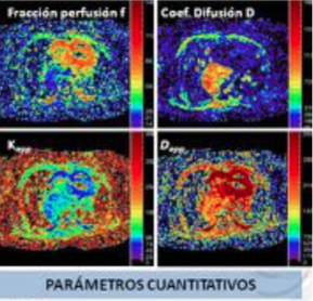
Existen varias características tumorales ultraestructurales que pueden ser evaluadas mediante varias métodos de imagen funcionales. De ellos, el más extendido en nuestro medio es la 18FDG-PET/TC para
la evaluación, predominantemente, del metabolismo tumoral (figura 1 y 2).
La aplicación de métodos de imagen basados en resonancia magnética (RM) en el tórax, aunque técnicamente difícil, están gradualmente más disponibles en la práctica clínica diaria. Además de la ausencia de radiación ionizante, una de sus mayores ventajas es la capacidad de evaluar diferentes características tisulares en un mismo protocolo, aportando una valoración integral multiparamétrica de lesiones tumorales y tejidos no tumorales.
Descargas
Citas
Le Bihan D (2013) Apparent diffusion coefficient and beyond: what diffusion MR imaging can tell us about tissue structure. Radiology 268:318–22.
Luna A, Sánchez-Gonzalez J, Caro P (2011) Diffusion-weighted imaging of the chest. Magn Reson Imaging Clin N Am 19:69–94.
Broncano J, Luna A, Sánchez-González J, et al. (2016) Functional MR Imaging in Chest Malignancies. Magn Reson Imaging Clin N Am 24:135–55.
Le Bihan D, Breton E, Lallemand D, et al. (1986) MR imaging of intravoxel incoherent motions: application to diffusion and perfusion in neurologic disorders. Radiology 161:401–7.
Takahara T, Kwee TC (2012) Low b-value diffusion-weighted imaging: emerging applications in the body. J Magn Reson Imaging 35:1266–73.
Le Bihan D, Breton E, Lallemand D, et al. (1988) Separation of diffusion and perfusion in intravoxel incoherent motion MR imaging. Radiology 168:497–505.
Rosenkrantz AB, Padhani AR, Chenevert TL, et al. (2015) Body diffusion kurtosis imaging: Basic principles, applications, and considerations for clinical practice. J Magn Reson Imaging n/a–n/a. 8. Runge VM, Clanton JA, Herzer WA, et al. (1984) Intravascular contrast agents suitable for magnetic resonance imaging. Radiology 153:171–6.
Buonaccorsi GA, Roberts C, Cheung S, et al. (2006) Comparison of the performance of tracer kinetic model-driven registration for dynamic contrast enhanced MRI using different models of contrast enhancement. Acad Radiol 13:1112–23.
Rijpkema M, Kaanders JH, Joosten FB, et al. (2001) Method for quantitative mapping of dynamic MRI contrast agent uptake in human tumors. J Magn Reson Imaging 14:457–63.
Carter BW, Benveniste MFK, Truong MT, Marom EM (2015) State of the Art: MR Imaging of Thymoma. Magn Reson Imaging Clin N Am 23:165–77.
Inaoka T, Takahashi K, Mineta M, et al. (2007) Thymic hyperplasia and thymus gland tumors: differentiation with chemical shift MR imaging. Radiology 243:869–76.
Inaoka T, Takahashi K, Iwata K, et al. (2005) Evaluation of normal fatty replacement of the thymus with chemical-shift MR imaging for identification of the normal thymus. J Magn Reson Imaging 22:341–6.
Ackman JB (2015) MR Imaging of Mediastinal Masses. Magn Reson Imaging Clin N Am 23:141–64.
Razek AA, Elmorsy A, Elshafey M, et al. (2009) Assessment of mediastinal tumors with diffusion-weighted single-shot echo-planar MRI. J Magn Reson Imaging 30:535–40.
Yabuuchi H, Matsuo Y, Abe K, et al. (2015) Anterior mediastinal solid tumours in adults: characterisation using dynamic contrast-enhanced MRI, diffusion-weighted MRI, and FDG-PET/CT. Clin Radiol 70:1289–98.
Duwe B V, Sterman DH, Musani AI (2005) Tumors of the mediastinum. Chest 128:2893–909. 18. Kosucu P, Tekinbas C, Erol M, et al. (2009) Mediastinal lymph nodes: assessment with diffusion-weighted MR imaging. J Magn Reson Imaging 30:292–7.
Shi HF, Feng Q, Qiang JW, et al. Utility of diffusion-weighted imaging in differentiating malignant from benign thyroid nodules with magnetic resonance imaging and pathologic correlation. J Comput Assist Tomogr 37:505–10.
Nomori H, Mori T, Ikeda K, et al. (2008) Diffusion-weighted magnetic resonance imaging can be used in place of positron emission tomography for N staging of non-small cell lung cancer with fewer false-positive results. J Thorac Cardiovasc Surg 135:816–22.
Tsuchida T, Morikawa M, Demura Y, et al. (2013) Imaging the early response to chemotherapy in advanced lung cancer with diffusion-weighted magnetic resonance imaging compared to fluorine-18 fluorodeoxyglucose positron emission tomography and computed tomography. J Magn Reson Imaging 38:80–8.
Ohno Y, Hatabu H, Takenaka D, et al. (2004) Metastases in mediastinal and hilar lymph nodes in patients with non-small cell lung cancer: quantitative and qualitative assessment with STIR turbo spin-echo MR imaging. Radiology 231:872–9.
Koyama H, Ohno Y, Nishio M, et al. (2014) Diffusion-weighted imaging vs STIR turbo SE imaging: capability for quantitative differentiation of small-cell lung cancer from non-small-cell lung cancer. Br J Radiol 87:20130307.
Matoba M, Tonami H, Kondou T, et al. (2007) Lung carcinoma: diffusion-weighted mr imaging--preliminary evaluation with apparent diffusion coefficient. Radiology 243:570–7.
Tanaka R, Nakazato Y, Horikoshi H, et al. Diffusion-weighted imaging and positron emission tomography in various cytological subtypes of primary lung adenocarcinoma. Clin Imaging 37:876–83. 26. Nomori H, Cong Y, Abe M, et al. (2015) Diffusion-weighted magnetic resonance imaging in preoperative assessment of non-small cell lung cancer. J Thorac Cardiovasc Surg 149:991–6.
Ohno Y, Nishio M, Koyama H, et al. (2014) Dynamic contrast-enhanced CT and MRI for pulmonary nodule assessment. AJR Am J Roentgenol 202:515–29.
Baysal T, Bulut T, Gökirmak M, et al. (2004) Diffusion-weighted MR imaging of pleural fluid: differentiation of transudative vs exudative pleural effusions. Eur Radiol 14:890–6.
Ohno Y, Koyama H, Yoshikawa T, et al. (2015) Lung Cancer Assessment Using MR Imaging: An Update. Magn Reson Imaging Clin N Am 23:231–244.
Ohno Y, Koyama H, Onishi Y, et al. (2008) Non-small cell lung cancer: whole-body MR examination for M-stage assessment--utility for whole-body diffusion-weighted imaging compared with integrated FDG PET/CT. Radiology 248:643–54.
Takenaka D, Ohno Y, Matsumoto K, et al. (2009) Detection of bone metastases in non-small cell lung cancer patients: comparison of whole-body diffusion-weighted imaging (DWI), whole-body MR imaging without and with DWI, whole-body FDG-PET/CT, and bone scintigraphy. J Magn Reson Imaging 30:298–308.
Yi CA, Shin KM, Lee KS, et al. (2008) Non-small cell lung cancer staging: efficacy comparison of integrated PET/CT versus 3.0-T whole-body MR imaging. Radiology 248:632–42.
Ohno Y, Nogami M, Higashino T, et al. (2005) Prognostic value of dynamic MR imaging for non-small-cell lung cancer patients after chemoradiotherapy. J Magn Reson Imaging 21:775–83.
Yabuuchi H, Hatakenaka M, Takayama K, et al. (2011) Non-Small Cell Lung Cancer: Detection of Early Response to Chemotherapy by Using Contrast-enhanced Dynamic and Diffusion-weighted MR Imaging. Radiology 261:598–604.
Figueiras RG, Padhani AR, Goh VJ, et al. (2011) Novel Oncologic Drugs: What They Do and How They Affect Images. Radiographics 31:2059–2091.
Razek AA, Elmorsy A, Elshafey M, et al. (2009) Assessment of mediastinal tumors with diffusion-weighted single-shot echo-planar MRI. J Magn Reson Imaging 30:535–40.
Van Rossum PSN, van Lier ALHMW, Lips IM, et al. (2015) Imaging of oesophageal cancer with FDG-PET/CT and MRI. Clin Radiol 70:81–95.
Chhabra A, Thakkar RS, Andreisek G, et al. (2013) Anatomic MR imaging and functional diffusion tensor imaging of peripheral nerve tumors and tumorlike conditions. AJNR Am J Neuroradiol 34:802–7.
Shin KE, Yi CA, Kim TS, et al. (2014) Diffusion-weighted MRI for distinguishing non-neoplastic cysts from solid masses in the mediastinum: problem-solving in mediastinal masses of indeterminate internal characteristics on CT. Eur Radiol 24:677–84.