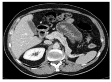
LESIONES HIPERVASCULARES PANCRÉATICAS
Palabras clave:
lesiones, hipervasculares, diagnóstico, diferencial, pancreas, pancreáticoResumen
1 Diagnóstico diferencial de las lesiones pancreáticas únicas o múltiples hipervasculares en el contexto de pacientes con antecedente oncológico de tumor primario hipervascular, fundamentalmente en el carcinoma renal. El problema clínico-radiológico que se plantea para distinguir entre una metástasis de carcinoma de células renales (fuerte tropismo para metastatizar en páncreas), y los mucho más frecuentes TNE.
2 Lesiones hipervasculares pancreáticas únicas vs múltiples en pacientes sin antecedentes oncológicos previos en el contexto de TNE único o Síndromes familiares (Men1, Sd VHL..)
3 Lesión hipervascular única en páncreas confirmada con AP como TNE y con comportamiento radiológico discretamente atípico en las pruebas de imagen.
Descargas
Citas
Kassabian A, Stein J, Jabbour N, Parsa K, Skinner D, Parekh D, et al. Renal cell carcinoma metastatic to the pancreas: a single-institution series and review of the literature. Urology. 2000 Aug 1;56(2):211–5.
Raman SP, Hruban RH, Cameron JL, Wolfgang CL, Fishman EK. Pancreatic imaging mimics: part 2, pancreatic neuroendocrine tumors and their mimics. AJR Am J Roentgenol. 2012 Aug;199(2):309–18. 3. Coakley FV, Hanley-Knutson K, Mongan J, Barajas R, Bucknor M, Qayyum A. Pancreatic imaging mimics: part 1, imaging mimics of pancreatic adenocarcinoma. AJR Am J Roentgenol. 2012 Aug;199(2):301–8.
Lewis RB, Lattin GE, Paal E. Pancreatic endocrine tumors: radiologic-clinicopathologic correlation. Radiographics. 2010 Oct;30(6):1445–64.
Xue H-D, Liu W, Xiao Y, Sun H, Wang X, Lei J, et al. Pancreatic and peri-pancreatic lesions mimic pancreatic islet cell tumor in multidetector computed tomography. Chin Med J. 2011 Jun;124(11):1720–5.
Manfredi R, Bonatti M, Mantovani W, Graziani R, Segala D, Capelli P, et al. Non-hyperfunctioning neuroendocrine tumours of the pancreas: MR imaging appearance and correlation with their biological behaviour. Eur Radiol. 2013 Nov;23(11):3029–39.
Scarsbrook AF, Thakker RV, Wass JAH, Gleeson FV, Phillips RR. Multiple endocrine neoplasia: spectrum of radiologic appearances and discussion of a multitechnique imaging approach. Radiographics. 2006 Apr;26(2):433–51.
Shi H, Zhao X, Miao F. Metastases to the Pancreas: Computed Tomography Imaging Spectrum and Clinical Features. Medicine (Baltimore) [Internet]. 2015 Jun 12 [cited 2016 Mar 16];94(23). Available from: http://www.ncbi.nlm.nih.gov/pmc/articles/PMC4616474/
Vincenzi M, Pasquotti G, Polverosi R, Pasquali C, Pomerri F. Imaging of pancreatic metastases from renal cell carcinoma. Cancer Imaging. 2014;14:5.
Leung RS, Biswas SV, Duncan M, Rankin S. Imaging Features of von Hippel–Lindau Disease. RadioGraphics. 2008 Jan 1;28(1):65–79.
Guan Z, Xu B, Wang R, Sun L, Tian J. Hyperaccumulation of (18)F-FDG in order to differentiate solid pseudopapillary tumors from adenocarcinomas and from neuroendocrine pancreatic tumors and review of the literature. Hell J Nucl Med. 2013 Aug;16(2):97–102.
Levy AD, Patel N, Dow N, Abbott RM, Miettinen M, Sobin LH. From the archives of the AFIP: abdominal neoplasms in patients with neurofibromatosis type 1: radiologic-pathologic correlation. Radiographics. 2005 Apr;25(2):455–80.
Kang TW, Kim SH, Lee J, Kim AY, Jang KM, Choi D, et al. Differentiation between pancreatic metastases from renal cell carcinoma and hypervascular neuroendocrine tumour: Use of relativepercentage washout value and its clinical implication. Eur J Radiol. 2015 Nov;84(11):2089–96.
Klein KA, Stephens DH, Welch TJ. CT characteristics of metastatic disease of the pancreas. Radiographics. 1998 Apr;18(2):369–78.
Bhavsar AS, Verma S, Lamba R, Lall CG, Koenigsknecht V, Rajesh A. Abdominal manifestations of neurologic disorders. Radiographics. 2013 Feb;33(1):135–53.


