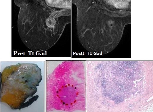
Estudio de la correlación radiopatológica de RM con diferentes modelos de respuesta en anatomía patológica en pacientes con neoadyuvancia por cáncer de mama localmente avanzado
Palabras clave:
neoadyuvancia, poster, seram, cáncer de mama, avanzado, correlación RM-AP, AP, anatomía patológicaResumen
Objetivo
• Revisar la correlación de los hallazgos en RM con diferentes modelos de valoración de respuesta en anatomía patológica de la pieza quirúrgica en pacientes con cáncer de mama localmente avanzado tratadas con neoadyuvancia en nuestro centro.
Material y método
- Realizamos un estudio descriptivo retrospectivo.
- Revisamos 55 pacientes con cáncer de mama localmente avanzado con tratamiento neoadyuvante previo a la cirugía entre enero de 2009 y febrero de 2016.
- En todas las pacientes se realizó:
- AP. Biopsia al diagnóstico y análisis de pieza quirúrgica tras tratamiento.
- RM pre y posttratamiento , previo a cirugía.
Descargas
Citas
Goldhirsch A, Wood WC, Coates AS, GelberRD, ThürlimannB, SennHJ. Strategies for subtypes–dealing with the diversity of breast cancer: highlights of the St. GallenInternational Expert Consensus on the Primary Therapy of Early Breast Cancer 2011. Ann Oncol. 2011;22:1736-47.
Tomida•K, Ishida M, UmedaT, Sakai S, Kawai Y, Mori T, Kubota Y, MekataE, Naka S, Abe H, Okabe H, TaniT. Magnetic resonance imaging shrinkage patterns following neoadjuvantchemotherapy for breast carcinomas with an emphasis on the radiopathologicalcorrelations.Mol ClinOncol. 2014 Sep;2(5):783-788. Epub2014 Jun 30 .
Kim TH, Kang DK, Yim H, Jung YS, Kim KS, Kang SY. •Magnetic resonance imaging patterns of tumorregression after neoadjuvantchemotherapy in breast cancer patients: correlation with pathological response grading system based on tumorcellularity.J ComputAssistTomogr. 2012 Mar-Apr;36(2):200-6. doi: 10.1097/RCT.0b013e318246abf3.
•Kim HJ, ImYH, Han BK, Choi N, Lee J, Kim JH, Choi YL, AhnJS, Nam SJ, Park YS, ChoeYH, KoYH, Yang JH. Accuracy of MRI for estimating residual tumorsize after neoadjuvantchemotherapy in locally advanced breast cancer: relation to response patterns on MRI. ActaOncol. 2007;46(7):996-1003.
Tozaki•M, Uno S, Kobayashi T, AibaK, Yoshida K, Takeyama H, ShioyaH, TabeiI, ToriumiY, Suzuki M, Kawakami M, Fukuda K. Histologic breast cancer extent after neoadjuvantchemotherapy: comparison with multidetector-row CT and dynamic MRI. RadiatMed. 2004 Jul-Aug;22(4):246-53.
Nakamura S, •KenjoH, NishioT, KazamaT, DoiO, Suzuki K.Efficacy of 3D-MR mammography for breast conserving surgery after neoadjuvantchemotherapy. Breast Cancer. 2002;9(1):15-9.
Nakamura S, •KenjoH, NishioT, KazamaT, Do O, Suzuki K. 3D-MR mammography-guided breast conserving surgery after neoadjuvantchemotherapy: clinical results and future perspectives with reference to FDG-PET. Breast Cancer. 2001;8(4):351-4.
Lakhani SR, Ellis IO, •SchnittSJ, Tan PH, Van de VijverMJ, eds. WHO classification of tumors of the breast.4th ed. Lyon:IARC;2012.
Symmans•WF, PeintingerF, HatzisC Rajan R, KuererH, Valero V, Assad L, PonieckaA, HennessyB,Green M, BudzarA, SingletaryE, HortobagyiGN, PusztaiL. Measurementof residual breastcancerburdento predictsurvivalafterneoadjuvantchemotherapy. J ClinOncol. 2007;25:4414-22.
Ogston•KN, Miller ID, Payne S, Hutcheon AW, Sarkar TK, Smith I, Schofield A, HeysSD. A new histological grading system to assess response of breast cancers to primary chemotherapy: prognostic significance and survival. Breast 2003;12:320-7.


