FACTORES RELACIONADOS CON LAS COMPLICACIONES TRAS BIOPSIA PULMONAR GUIADA POR TOMOGRAFÍA COMPUTARIZADA (TC):
ANÁLISIS RESTROSPECTIVO DE CASOS Y CONTROLES DE 112 PROCEDIMIENTOS
Palabras clave:
BIOPSIA PULMONAR GUIADA, POSTER, SERAM, COMPLICACIONES, TCResumen
OBJETIVOS
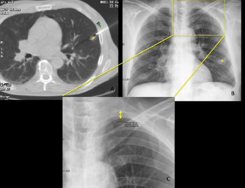
La biopsia con aguja gruesa (BAG) percutánea guiada por tomografía computarizada se utiliza actualmente en la vía de diagnóstico de las lesiones pulmonares. La etiología de tales lesiones pulmonares puede variar de benigna a maligna lo que cambia el tratamiento necesario. Por ello es necesario establecer un diagnóstico definitivo.
Sin embargo, no podemos olvidar que la BAG pulmonar guiada por TC es un procedimiento invasivo que tiene complicaciones.
MATERIAL Y MÉTODO
En este estudio retrospectivo de casos y controles apareados , revisamos la base de datos del Departamento de Radiología Torácica. Esta base de datos se actualiza cada vez que hay un procedimiento intervencionista de tórax, registrando dicho procedimiento y sus características más relevantes, incluyendo si se utilizó o no suero salino al extraer la aguja coaxial. Esta técnica fue utilizada por primera vez en 2013 por uno de los radiólogos torácicos, al leer en la literatura existente que era un técnica beneficiosa sin riesgos añadidos, y desde entonces los otros dos radiólogos que conforman el área también la incorporaron a la
rutina intervencionista en BAG guida por TC, siguiendo sus propios criterios. Finalmente, la técnica se generalizó.
Descargas
Citas
• Klein JS, Salomon G, Stewart EA. Transthoracic needle biopsy with a coaxially placed 20 gauge automated cutting needle, results in 122 patients. Radiology 1995;198:715-720.
• Li Y, Du Y, Yang HF, et al . CT guided percutaneous core needle biopsy for small (≤20 mm) pulmonary lesions. ClinRadiol 2013;68:e43-8.
• YaffeD,ShitritD,GottfriedM, etal .Ipsilateralopposite sideaspirationin resistant pneumothorax after CT image guided lung biopsy: complementary role after simple needle aspiration. Chest 2013;144:947-51.
• Yeow KM, Su IH, Pan KT, et al. Risk factors of pneumothorax and bleeding: multivariate analysis of 660 CT guided coaxial cutting needle lung biopsies. Chest 2004;126:748-54.
• Laurent F, Latrabe V, Vergier B, et al . CT guided transthoracic needle biopsy of pulmonary nodules smaller than 20 mm: results with an automated 20 gauge coaxial cutting needle. ClinRadiol 2000;55:281-7.
• Montaudon M, Latrabe V, Pariente A, et al . Factors influencing accuracy of CT guided percutaneous biopsies of pulmonary lesions. EurRadiol 2004;14:1234-40.
• Yamagami T, Nakamura T, Iida S, et al . Management of pneumothorax after percutaneous CT guided lung biopsy. Chest 2002;121:1159-64.
• Yildirim E, Kirbas I, Harman A, et al. CT guided cutting needle lung biopsy using modified coaxial technique: factors effecting risk of complications. Eur J Radiol 2009;70:5760.
• Li, Y., Du, Y., Luo , T., Yang, H., Yu, J., Xu , X., Zheng , H. and Li, B..Usefulness of normal saline for sealing the needle track after CT guided lung biopsy. ClinicalRadiology 2015;70(11), pp.1192-1197.
• Billich , C., Muche , R., Brenner, G., Schmidt, S., Krüger, S., Brambs , H. and Pauls, S. CT guided lung biopsy: incidence of pneumothorax after instillation of NaCl into the biopsy track. European Radiology 2008;18(6), pp.1146-1152.


