COLECISTITIS AGUDA, SER O NO SER: ÉSA ES LA CUESTIÓN.
CONTROVERSIAS EN LA CONFIANZA DIAGNÓSTICA DE LA COLECISTITIS AGUDA Y ALTERNATIVAS EN EL MANEJO DE LA IMAGEN.
Palabras clave:
COLECISTITIS AGUDA, poster, seramResumen
Objetivos Docentes
• Revisión de las últimas actualizaciones en el manejo diagnóstico de la colecistitis aguda
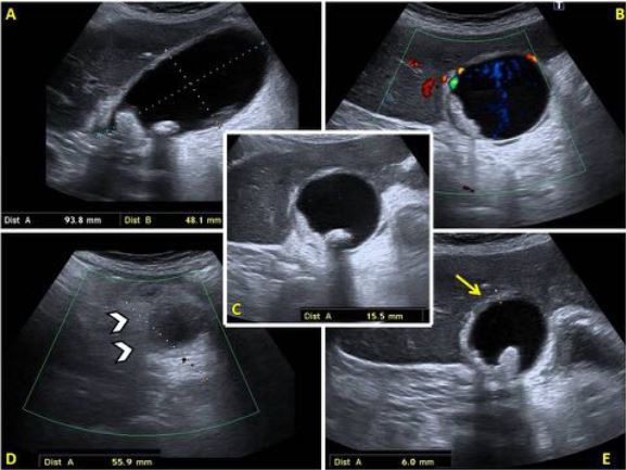
• Análisis de los signos diagnósticos por imagen clásicos y su rentabilidad y controversia, tanto de forma aislada como en combinación.
• Valoración de las alternativas en el proceso diagnóstico, enfatizando el uso del TC y signos precoces por ecografía.
Revisión del tema
INTRODUCCIÓN:
El dolor agudo focalizado en hipocondrio derecho es un síntoma frecuente en el contexto de la urgencia, así como ocasionalmente en pacientes graves hospitalizados por otras causas. Uno de los principales diagnósticos a descartar es la colecistitis aguda (CA), la cual debe ser manejada de manera urgente, por sus probables complicaciones y potencial de mortalidad [1].
Descargas
Citas
Connor O, Maher M. Imaging in cholecystitis. AJR 2011; 196: 367 – 374.
Ann C, Park S, Ko S, Par MS, Kim M, Kim KW. Usefulness of the tensile gallbladder fundus sign in the diagnosis of early acute cholecystitis. AJR 2013; 201: 340 – 346.
Trowbridge RL, Rutkowski NK, Shojania KG. Does hispatient hace acutecholecystitis? JAMA 2003; 289: 80 – 86.
Yokoe M, Takada T, Strasberg S, Solomkin J, Mayumi T, Gomi H et al. TG13 diagnostic criteria and severity grading of acute cholecystitis. J HepatobiliaryPancreaticSci 2013; 20: 35 – 46.
Yokoe M, Takada T, Strasberg S, Solomkin J, Mayumi T, Gomi H et al. New diagnosticcriteria and severityassessment of acutecholecystitis in revised Tokio guidelines. J HepatobiliaryPancreaticSci 2012; 19: 578 – 85.
Hirota M, Takada T, Kawarada Y, Nimura Y, Miura F, Hirata K, et al. Diagnostic criteria and severity assessment of acute cholecystitis: Tokyo Guidelines. J Hepatobiliary Pancreat Surg. 2007; 14(1): 78 – 82.
Gruber PJ, Silverman RA, Gottesfeld S, Dlaster E. Presence of fever and leukocytosis in acutecholecystitis. Ann EmergMed. 1996; 28: 273 – 7.
Yarmish G, Smith M, Rosen M, Baker M, Blake M, Cash B, et al. ACR AppropiatenessCriteriaRightUpperQuadrantPain. J Am CollRadiol 2014; 11: 316 – 322.
Hwang H, Marsh I, Doyle J. Does ultrasonography accurately diagnose acute cholecystitis?
Improving diagnostic accuracy based on a review at a regional hospital. Can J Surg 2014; 57 (3): 162 – 168.
Kiewer JJ, Leeuwenburgh MM, Bipat S, Bossuyt PM, Stoker J, Boermeester MA. A systematicreview and meta-analysis of diagnostic performance of imaging in acutecholecystitis. Radiology 2012; 264: 708 – 20.
Alobaidi M, Gupta R, Jafri SZ, Fink .Benner DM. Current trends in imaging evaluation of acute cholecystitis. EmergRadiol 2004; 10: 256 – 8.
Shakespear J, Shaaban A, Rezvani M. CT findings f acutecholecystitis and itscomplications. AJR 2010; 194: 1523 – 29.
Vriesman A, Engelbrecht M, Smithuis R, Puylaert J. Diffuse gallbladder wall thickening: differential diagnosis. AJR 2007; 188: 495 – 501.
Cohan RH, Mahony BS, Bowie JD, Cooper C, Baker ME, Illescas FF. Striatedintramuralgallbladderlucencieson US studies: predictors of acutecholecystitis. Radiology 1987; 164: 31 – 5.
Jeffrey RB Jr, Nino – Murcia M, ralls PW, Jain KA, Davidson HC. Color Dopplersonography of thecysticartery: comparison of normal controls and patientswithacutecholecystitis. J UltrasoundMed. 1995; 14: 33 – 6.
Forsberg L, ANdersson R, Hederstrom E, Tranberg KG. Ultrasonography and gallbladderperforation in acutecholecystitis. Acta Radiol. 1988; 29: 203 – 5.
Aydin C, et al, en prognostic parameters for the prediction of acute gangrenous cholecystitis. J Hepatobiliary pancreat surg 2006: 13: 155 – 159
Teefey S, Dahiya N, Middleton W, Bajaj S, Dahiya N, Ylagan L, et al. Acute cholecystitis: do sonographic findings and WBC count predict gangrenous changes?. AJR 2013; 200: 363 – 369.
Fidler J, Paulson E, Layfield L. CT evaluation of acute cholecystitis. Findings and usefullness in diagnosis. AJR 1996; 166: 1085 – 88.
Boork O, Kane R, Tyagu G, Siewert B, Kruskal J. Lessons learned from quality assurance: errors in the diagnosis of acute cholecystitis on ultrasound and CT. AJR 2001; 196: 597 – 604.
Kim YK, Kwak HS, Kim CS et al. CT findings of mild forms or early manifestations of acute cholecystitis. Clin Imaging 2009; 33: 274 – 280
Aoun N, Smayra T, Haddad – Zebouni S, Slaba S, Ghossain M, Atalla N. Acute cholecystitis using computed tomography: usefulness of the trabeculation of the peri – cholecystic adipose tissue. J Radiol 1999; 80 (6): 575 – 8.
Soyer P, Hoeffel C, Dohan A, Gayat E, Eveno C; Malgras B et al. Acute cholecystitis: quantitiative and qualitative evaluation with 64 – section helical CT. Acta Radiol 2013; 54 (5): 477– 86