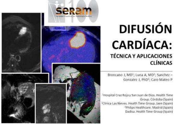
Difusión cardíaca:
Técnica y aplicaciones clínicas.
Palabras clave:
Difusión cardíaca, poster, seram, (Diffusion Weighted Imaging, DWIResumen
Objetivos Docentes
Objetivos:
1. Analizar los ajustes técnicos necesarios para la realización de imagen potenciada en difusión en el corazón.
2. Revisar las potenciales aplicaciones clínicas de la difusión cardíaca.
Revisión del tema
1. Introducción
Gracias a los avances recientes en la imagen funcional y molecular las secuencias de RM han consolidado una mejora sustancial en la caracterización de tejidos normales y patológicos. La difusión por RM (Diffusion Weighted Imaging; DWI) se ha convertido en una herramienta poderosa desde el punto de vista diagnóstico, no sólo para la diferenciación entre lesiones malginas y benignas, sino como biomarcador oncológico con importantes implicaciones pronósticas en diferentes partes de nuestra anatomía.
Descargas
Citas
Le Bihan D (2013) Apparent diffusion coefficient and beyond: what diffusion MR imaging can tell us about tissue structure. Radiology 268:318–22.
Luna A, Sánchez-Gonzalez J, Caro P (2011) Diffusion-weighted imaging of the chest. Magn Reson Imaging Clin N Am 19:69–94.
Choi JS, Kim M-J, Chung YE, et al. (2013) Comparison of breathhold, navigator-triggered, and free-breathing diffusion-weighted MRI for focal hepatic lesions. J Magn Reson Imaging 38:109–18.
Laissy J-P, Gaxotte V, Ironde-Laissy E, et al. (2013) Cardiac diffusion-weighted MR imaging in recent, subacute, and chronic myocardial infarction: a pilot study. J Magn Reson Imaging 38:1377–87.
Broncano J, Luna A, Sánchez-González J, et al. (2016) Functional MR Imaging in Chest Malignancies. Magn Reson Imaging Clin N Am 24:135–55.
Padhani AR, Liu G, Koh DM, et al. (2009) Diffusion-weighted magnetic resonance imaging as a cancer biomarker: consensus and recommendations. Neoplasia 11:102–25.
Le Bihan D, Breton E, Lallemand D, et al. (1986) MR imaging of intravoxel incoherent motions: application to diffusion and perfusion in neurologic disorders. Radiology 161:401–7.
Le Bihan D, Breton E, Lallemand D, et al. (1988) Separation of diffusion and perfusion in intravoxel incoherent motion MR imaging. Radiology 168:497–505.
Eitel I, Friedrich MG (2011) T2-weighted cardiovascular magnetic resonance in acute cardiac disease. J Cardiovasc Magn Reson 13:13.
Kociemba A, Pyda M, Katulska K, et al. (2013) Comparison of diffusion-weighted with T2-weighted imaging for detection of edema in acute myocardial infarction. J Cardiovasc Magn Reson 15:90.
Lloyd-Jones D, Adams RJ, Brown TM, et al. (2010) Executive summary: heart disease and stroke statistics--2010 update: a report from the American Heart Association. Circulation 121:948–54.
Alpert JS, Thygesen K, Antman E, Bassand JP (2000) Myocardial infarction redefined--a consensus document of The Joint European Society of Cardiology/American College of Cardiology Committee for the redefinition of myocardial infarction. J Am Coll Cardiol 36:959–69.
Thygesen K, Alpert JS, Jaffe AS, et al. (2012) Third universal definition of myocardial infarction. Circulation 126:2020–35.
Renker M, Baumann S, Rier J, et al. (2015) Imaging coronary artery disease and the myocardial ischemic cascade: clinical principles and scope. Radiol Clin North Am 53:261–9.
Rajiah P, Desai MY, Kwon D, Flamm SD (2013) MR Imaging of Myocardial Infarction. RadioGraphics 33:1383–1412.
Friedrich MG (2010) Myocardial edema--a new clinical entity? Nat Rev Cardiol 7:292–6.
von Knobelsdorff-Brenkenhoff F, Schulz-Menger J (2012) Cardiovascular magnetic resonance imaging in ischemic heart disease. J Magn Reson Imaging 36:20–38.
Laissy JP, Serfaty JM, Messika-Zeitoun D, et al. (2009) [Cardiac diffusion MRI of recent and chronic myocardial infarction: preliminary results]. J Radiol 90:481–4.
Friedrich MG, Sechtem U, Schulz-Menger J, et al. (2009) Cardiovascular magnetic resonance in myocarditis: A JACC White Paper. J Am Coll Cardiol 53:1475–87.
Cooper LT (2009) Myocarditis. N Engl J Med 360:1526–38.
Liu PP, Mason JW (2001) Advances in the Understanding of Myocarditis. Circulation 104:1076–1082.
Potet J, Rahmouni A, Mayer J, et al. (2013) Detection of myocardial edema with low-b-value diffusion-weighted echo-planar imaging sequence in patients with acute myocarditis. Radiology 269:362–9.
Rapacchi S, Wen H, Viallon M, et al. (2011) Low b-value diffusion-weighted cardiac magnetic resonance imaging: initial results in humans using an optimal time-window imaging approach. Invest Radiol 46:751–8.
Bogaert J, Francone M (2013) Pericardial disease: value of CT and MR imaging. Radiology 267:340–56.
Cosyns B, Plein S, Nihoyanopoulos P, et al. (2015) European Association of Cardiovascular Imaging (EACVI) position paper: Multimodality imaging in pericardial disease. Eur Heart J Cardiovasc Imaging 16:12–31.
Bock JS, Benitez RM (2012) Blunt cardiac injury. Cardiol Clin 30:545–55. doi: 10.1016/j.ccl.2012.07.001
Restrepo CS, Vargas D, Ocazionez D, et al. (2013) Primary pericardial tumors. Radiographics 33:1613–30.
Motwani M, Kidambi A, Herzog BA, et al. (2013) MR imaging of cardiac tumors and masses: a review of methods and clinical applications. Radiology 268:26-43.
Yared K, Baggish AL, Picard MH, et al. (2010) Multimodality imaging of pericardial diseases. JACC Cardiovasc Imaging 3:650–60.
Gutberlet M, Spors B, Thoma T, et al. (2008) Suspected chronic myocarditis at cardiac MR: diagnostic accuracy and association with immunohistologically detected inflammation and viral persistence. Radiology 246:401–9.
Ohno Y, Koyama H, Yoshikawa T, et al. (2012) Diffusion-weighted MRI versus 18F-FDG PET/CT: performance as predictors of tumor treatment response and patient survival in patients with non-small cell lung cancer receiving chemoradiotherapy. AJR Am J Roentgenol 198:75–82.
Ohno Y, Sugimura K, Hatabu H (2002) MR imaging of lung cancer. Eur J Radiol 44:172–81.
Okuma T, Matsuoka T, Yamamoto a, et al. (2009) Assessment of early treatment response after CT-guided radiofrequency ablation of unresectable lung tumours by diffusion-weighted MRI: a pilot study. Br J Radiol 82:989–94.