Utilidad de la RM-difusión, tensor de difusión (DTI) y la tractografía en la valoración de la patología medular.
Bases físicas y aplicaciones clínicas.
Palabras clave:
patología medular, poster, seramResumen
Objetivos Docentes
Revisar las bases físicas de la secuencia de difusión y del tensor de difusión (DTI) así como los ajustes técnicos necesarios para su aplicación en el cordón medular
Mostrar las principales aplicaciones clínicas para la valoración de la patología del cordón medular a través de distintos escenarios clínicos.
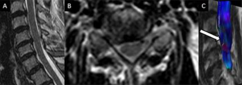
Revisión del tema
1.- Introducción
Las secuencias potenciadas en difusión (DWI) son capaces de detectar el movimiento de las moléculas de agua en un medio biológico. Dichas moléculas experimentan, en condiciones normales un desplazamiento a lo largo del espacio transcurrido un determinado periodo de tiempo. Por lo tanto, dichas secuencias no sólo permiten valorar cualitativamente dicho grado de movimiento sino también cuantificarlo aportando de esta forma información anatómica y funcional de los tejidos normales y patológicos.
Descargas
Citas
Sundgren PC, Dong Q, Gómez-Hassan D, Mukherji SK, Maly P, Welsh R. Diffusion tensor imaging of the brain: review of clinical applications. Neuroradiology [Internet]. 2004;46(5):339–50.
Barakat N, Mohamed FB, Hunter LN, Shah P, Faro SH, Samdani AF, et al. Diffusion tensor imaging of the normal pediatric spinal cord using an inner field of view echo-planar imaging sequence. Am J Neuroradiol. 2012;33(6):1127–33.
Vargas MI, Delavelle J, Kohler R, Becker CD, Lovblad K. Brain and spine MRI artifacts at 3 Tesla. J Neuroradiol. 2009;36(2):74–81.
Park EH, Lee YH, Jeong E-K, Roh YH, Suh J-S. Diffusion tensor imaging focusing on lower cervical spinal cord using 2D reduced FOV interleaved multislice single-shot diffusion-weighted echo-planar imaging: comparison with conventional single-shot diffusion-weighted echo-planar imaging. Magn Reson Imaging [Internet]. Elsevier Inc.; 2015;33(4):401–6.
Raya JG, Dietrich O, Birkenmaier C, Sommer J, Reiser MF, Baur-Melnyk A. Feasibility of a RARE-based sequence for quantitative diffusion-weighted MRI of the spine. Eur Radiol. 2007;17(11):2872–9.
El Maati AA a., Chalabi N. Diffusion tensor tractography as a supplementary tool to conventional MRI for evaluating patients with myelopathy. Egypt J Radiol Nucl Med [Internet]. Elsevier B.V.; 2014;45(4):1223–31.
Ducreux D, Fillard P, Facon D, Ozanne A, Lepeintre J-F, Renoux J, et al. Diffusion tensor magnetic resonance imaging and fiber tracking in spinal cord lesions: current and future indications. Neuroimaging Clin N Am [Internet]. 2007 Mar [cited 2013 Jul 31];17(1):137–47.
Setzer M, Murtagh RD, Murtagh FR, Eleraky M, Jain S, Marquardt G, et al. Diffusion tensor imaging tractography in patients with intramedullary tumors: comparison with intraoperative findings and value for prediction of tumor resectability. J Neurosurg Spine [Internet]. 2010 Sep [cited 2013 Aug 2];13(3):371–80.
Urger E, Debellis MD, Hooper SR, Woolley DP, Chen S, Provenzale JM. Influence of analysis technique on measurement of diffusion tensor imaging parameters. AJR Am J Roentgenol [Internet]. 2013 May [cited 2013 Jul 31];200(5):W510–7.
Bosemani T, Tekes A. Focal spinal cord lesions in children?: A practical approach. Appl Radiol. 2012;41(04 April).
Meier R, Thuermel K, Noel PB, Moog P, Sievert M, Ahari C, et al. Synovitis in patients with early inflammatory arthritis monitored with quantitative analysis of dynamic contrast-enhanced optical imaging and MR imaging. Radiology [Internet]. 2014;270(1):176–85.
Thurnher MM, Cartes-Zumelzu F, Mueller-Mang C. Demyelinating and infectious diseases of the spinal cord. Neuroimaging Clin N Am [Internet]. 2007 Mar [cited 2013 Jul 31];17(1):37–55.
Lee JW, Park KS, Kim JH, Choi J-Y, Hong SH, Park S-H, et al. Diffusion tensor imaging in idiopathic acute transverse myelitis. AJR Am J Roentgenol [Internet]. 2008 Aug [cited 2013 Jul 31];191(2):W52–7.
Vargas MI, Delavelle J, Jlassi H, Rilliet B, Viallon M, Becker CD, et al. Clinical applications of diffusion tensor tractography of the spinal cord. Neuroradiology [Internet]. 2008 Jan [cited 2013 Jul 31];50(1):25–9.
Hayes LL, Jones R a, Palasis S, Aguilera D, Porter D a. Drop metastases to the pediatric spine revealed with diffusion-weighted MR imaging. Pediatr Radiol [Internet]. 2012;42(8):1009–13.
Filippi CG, Andrews T, Gonyea J V, Linnell G, Cauley K a. Magnetic resonance diffusion tensor imaging and tractography of the lower spinal cord: application to diastematomyelia and tethered cord. Eur Radiol [Internet]. 2010 Oct [cited 2013 Jul 31];20(9):2194–9.
Hori M, Tsutsumi S, Yasumoto Y, Ito M, Suzuki M, Tanaka FS, et al. Cervical spondylosis: Evaluation of microstructural changes in spinal cord white matter and gray matter by diffusional kurtosis imaging. Magn Reson Imaging [Internet]. The Authors; 2014;32(5):428–32.
Lindberg PG, Sanchez K, Ozcan F, Rannou F, Poiraudeau S, Feydy A, et al. Correlation of force control with regional spinal DTI in patients with cervical spondylosis without signs of spinal cord injury on conventional MRI. Eur Radiol [Internet]. European Radiology; 2015;
Wang KY, Idowu O, Thompson CB, Orman G, Myers C, Riley LH, et al. Tract-Specific Diffusion Tensor Imaging in Cervical Spondylotic Myelopathy Before and After Decompressive Spinal Surgery: Preliminary Results. Clin Neuroradiol [Internet]. 2015;
Oakden W, Kwiecien JM, O’Reilly M a., Dabrowski W, Whyne C, Finkelstein J, et al. Quantitative MRI in a non-surgical model of cervical spinal cord injury. NMR Biomed [Internet]. 2015;(April)