El papel de la Radiología en el estudio anatomo-patológico del cáncer de pulmón:
De la PAAF a la BAG y al arpón pulmonar.
Palabras clave:
cáncer de pulmón, poster, seram, PAAF, BAG, arpón pulmonar, CPNM, CPMResumen
Objetivos Docentes
1. Revisar las técnicas mínimamente invasivas empleadas en Radiología (PAAF, BAG y arpón pulmonar) para la obtención de material para estudio anatomo-patológico en cáncer de pulmón.
2. Revisar sus indicaciones, contraindicaciones, complicaciones y rentabilidad diagnóstica.
Revisión del tema
INTRODUCCION
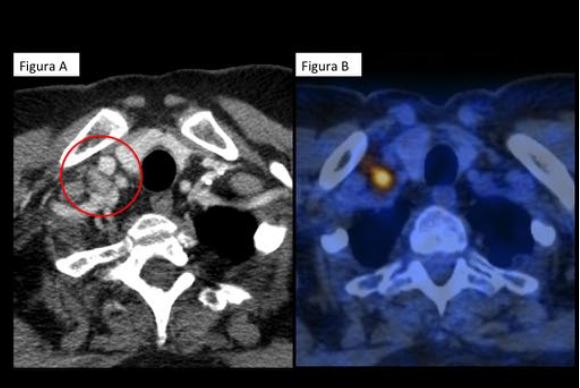
La obtención de muestras para estudio anatomopatológico en cáncer de pulmón con técnicas mínimamente invasivas es práctica habitual en los Servicios de Radiología. Es habitual realizar punciones transtorácicas guiadas con un método de imágen (generalmente la TC) y tradicionalmente su objetivo ha sido (1) la confirmación diagnóstica de carcinoma broncogénico y (2) la diferenciación entre carcinoma microcítico (CPM) y no microcítico (CPNM) para la elección de tratamiento. Posteriormente se añadió la subclasificación histológica del CPNM (fundamentalmente adenocarcinoma, carcinoma escamoso y epidermoide y carcinoma de células grandes). La punción aspiración con aguja fina (PAAF) permite diferenciar entre CPNM y CPM y entre los subtipos histológicos del CPNM.
Descargas
Citas
Beslic S, Zukic F, Milisic S. Percutaneous transthoracic CT guided biopsies of lung lesions; fine needle aspiration biopsy versus core biopsy. Radiol Oncol. 2012 Mar;46(1):19–22.
Chen Y-R, Yeow K-M, Lee J-Y, Su I-H, Chu S-Y, Lee C-H, et al. CT-guided hook wire localization of subpleural lung lesions for video-assisted thoracoscopic surgery (VATS). J Formos Med Assoc. 2007 Nov;106(11):911–8.
Hanauer M, Perentes JY, Krueger T, Ris H-B, Bize P, Schmidt S, et al. Pre-operative localization of solitary pulmonary nodules with computed tomography-guided hook wire: report of 181 patients. J Cardiothorac Surg. 2016;11(1):5.
Ichinose J, Kohno T, Fujimori S, Harano T, Suzuki S. Efficacy and complications of computed tomography-guided hook wire localization. The Annals of Thoracic Surgery. 2013 Oct;96(4):1203–8.
Isaka T, Ito H, Yokose T, Kondo T, Nagata M, Nishii T, et al. Prediction of lung tumor palpability using high-resolution computed tomography. Asian Cardiovascular and Thoracic Annals. SAGE Publications; 2016 Jan;24(1):23–9.
Kim L, Tsao MS. Tumour tissue sampling for lung cancer management in the era of personalised therapy: what is good enough for molecular testing? European Respiratory Journal. 2014 Sep 30;44(4):1011–22.
Lazarus DR, Ost DE. How and when to use genetic markers for nonsmall cell lung cancer. Current Opinion in Pulmonary Medicine. 2013 Jul;19(4):331–9.
Molins L, Mauri E, Sánchez M, Fibla JJ, Gimferrer JM, Arguis P, et al. Localización de nódulos pulmonares con arpón guiado por tomografía axial computarizada previa a la resección videotoracoscópica. Experiencia en 52 casos. Cirugía Española. 2013 Mar;91(3):184–8.
Ofiara LM, Navasakulpong A, Beaudoin S, Gonzalez AV. Optimizing tissue sampling for the diagnosis, subtyping, and molecular analysis of lung cancer. Front Oncol. Frontiers; 2014;4(Suppl):253.
Otto S, Mensel B, Friedrich N, Schäfer S, Mahlke C, Bernstorff von W, et al. Predictors of Technical Success and Rate of Complications of Image-Guided Percutaneous Transthoracic Lung Needle Biopsy of Pulmonary Tumors. Schmidt RL, editor. PLoS ONE. Public Library of Science; 2015 Apr 9;10(4):e0124947–9.
Patel IJ, Davidson JC, Nikolic B, Salazar GM, Schwartzberg MS, Walker TG, et al. Consensus Guidelines for Periprocedural Management of Coagulation Status and Hemostasis Risk in Percutaneous Image-guided Interventions. J Vasc Interv Radiol. Elsevier Inc; 2012 Jun 1;23(6):727–36.
Pirker R, Herth FJF, Kerr KM, Filipits M, Taron M, Gandara D, et al. Consensus for EGFR mutation testing in non-small cell lung cancer: results from a European workshop. Elsevier; 2010. pp. 1706–13.
Saito H, Minamiya Y, Matsuzaki I, Tozawa K, Taguchi K, Nakagawa T, et al. Indication for preoperative localization of small peripheral pulmonary nodules in thoracoscopic surgery. The Journal of Thoracic and Cardiovascular Surgery. Elsevier; 2002 Dec;124(6):1198–202.
Suzuki K, Nagai K, Yoshida J. Video-assisted thoracoscopic surgery for small indeterminate pulmonary nodules: indications for preoperative marking. Chest. 1999;115(2):563–8.
Tamura M, Oda M, Fujimori H, Shimizu Y, Matsumoto I, Watanabe G. New indication for preoperative marking of small peripheral pulmonary nodules in thoracoscopic surgery. Interactive CardioVascular and Thoracic Surgery. 2010 Oct 20;11(5):590–3.
Winokur R, Pua B, Sullivan B, Madoff D. Percutaneous Lung Biopsy: Technique, Efficacy, and Complications. Seminars in Interventional Radiology. 2013 May 28;30(02):121–7.
Wu K, House L, Liu W, Cho WCS. Personalized Targeted Therapy for Lung Cancer. IJMS. 2012 Dec;13(12):11471–96.