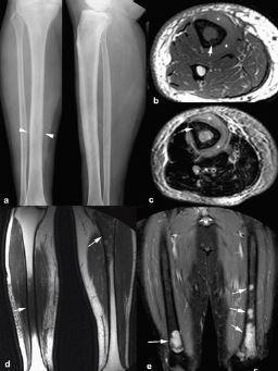
RM de los linfomas musculoesqueléticos primarios:
claves diagnósticas
Palabras clave:
linfomas musculoesqueléticos primarios, poster, seramResumen
Objetivos Docentes
• Revisar las manifestaciones en RM de los linfomas musculoesqueléticos primarios.
• Presentar los hallazgos semiológicos más útiles para sugerir el diagnóstico
Revisión del tema
La afectación ósea, muscular, del tejido celular subcutáneo y de la piel de los linfomas suele sersecundaria por diseminación hematógena o extensión por continuidad desde los ganglios afectados por el tumor.
La afectación musculoesquelética primaria de los linfomas es rara, supone el 0,3% de los linfomas de Hodgkin y el 1,5% de los no-Hodgkin.
Descargas
Citas
Chun CW, Jee WH, Park HJ, et al. MRI features of skeletal muscle lymphoma. AJR Am J Roentgenol 2010;195:1355-60.
Heyning FH, Kroon HM, Hogendoorn PC, Taminiau AH, van der Woude HJ. MR imaging characteristics in primary lymphoma of bone with emphasis on non-aggressive appearance.
Skeletal Radiol 200;36:937-44.
Hwang S. Imaging of lymphoma of the musculoskeletal system. Magn Reson Imaging Clin N Am 2010;18:75-93.
Kang BS, Choi SH, Cha HJ, Jet al. Subcutaneous panniculitis-like T-cell lymphoma: US and CT findings in three patients. Skeletal Radiol 2007 Jun;36 Suppl 1:S67-71.
Levine BD, Seeger LL, James AW, Motamedi K. Subcutaneous panniculitis-like T-cell lymphoma: MRI features and literature review. Skeletal Radiol 2014;43:1307-11.
Mengiardi B, Honegger H, Hodler J, Exner UG, Csherhati MD, Brühlmann W. Primary lymphoma of bone: MRI and CT characteristics during and after successful treatment. AJR Am J Roentgenol 2005;184:185-92.
Surov A. Imaging findings of skeletal muscle lymphoma. Clin Imaging 2014;38:594-8.


