La radiología de la columna vertebral mediante TC y RM hecha fácil
Palabras clave:
columna vertebral, poster, seramResumen
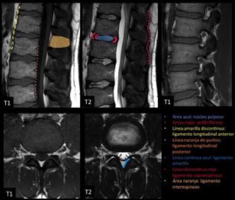
Objetivos Docentes
a. Repasar la anatomía de la columna vertebral.
b. Conocer las claves para la correcta valoración de los estudios de TC y RM de la columna.
c. Revisar la patología más frecuente de causa traumática y no traumática a través de casos observados en nuestro centro.
Revisión del tema
El estudio de la columna vertebral es complejo y supone a menudo un reto diagnóstico para el radiólogo.
Mediante esta revisión se pretende simplificar el abordaje de la misma y exponer las claves radiológicas para reconocer en los estudios de TC y RM las patologías que más frecuentemente afectan a la columna vertebral, tanto de etiología traumática como no traumática.
Descargas
Citas
Jeffrey S. Ross, Kevin R. Moore, Bryson Borg, Julia Crim, Lubdha M. Shah. Diagnostic Imaging: Spine. Second edition.
P. M. Parizel, T. van der Zijden, S. Gaudino et al. Trauma of the spine and spinal cord: imaging strategies. Eur Spine J (2010) 19 (Suppl 1):S8–S17.
Alexander R. Vaccaro, Ronald A. Lehman, Jr., R. John Hurlbert, Paul A. Anderson et al. A New Classification of Thoracolumbar Injuries. The Importance of Injury Morphology, the Integrity of the Posterior Ligamentous Complex, and Neurologic Status. SPINE Volume 30, Number 20, pp 2325–2333. 2005.
Roy Riascos, Eliana Bonfante, Claudia Cotes, Mary Guirguis et al. Imaging of AtlantoOccipital and Atlantoaxial Traumatic Injuries: What the Radiologist Needs to Know. RadioGraphics 2015; 35:21212134.
Christopher D. Chaput., Jonathan Walgama, Erick Torres, et al. Defining and Detecting Missed Ligamentous Injuries of the Occipitocervical Complex. SPINE Volume 36, Number 9, pp
2011.
David F. Fardon, Alan L. Williams, Edward J. Dohring, et al. Lumbar disc nomenclature: versión 2.0. Recommendations of the combined task forces of the North American Spine Society, the American Society of Spine Radiology and the American Society of Neuroradiology. The Spine Journal 14(2014) 25252545.
David Malfair, Douglas P. Beall. Imaging the Degenerative Diseases of the Lumbar Spine. Magnetic Resonance Imaging Clinics of North America Vol. 15, Issue 2, p221–238.
Amir M. Khan, Federico Girardi. Spinal lumbar synovial cysts. Diagnosis and management challenge. Euroean Spine Journal (2006) 15:11761182.
Nadja Mamisch, Martin Brumann, Juerg Hodler et al. Radiologic Criteria for the Diagnosis of Spinal Stenosis:Results of a Delphi Survey. Radiology: Volume 264: Number 1—July 2012.
Yusuhn Kang, Joon Woo Lee, Young Hwan Koh et al. New MRI Grading System for the Cervical Canal Stenosis. AJR 2011; 197:W134–W140.
Johann Steurer, Simon Roner, Ralph Gnannt et al. Quantitative radiologic criteria for the diagnosis of lumbar spinal stenosis: a systematic literatura review. BMC Musculoskeletal
Disorders 2011, 12:175.
Mathieu H. Rodallec, Antoine Feydy, Frédérique Larousserie et al. Diagnostic Imaging of Solitary Tumors of the Spine: What to Do and Say. RadioGraphics 2008; 28:1019–1041
Justin Alexander, Adam Meir, Nikitas Vrodos and Yun-Hom Yau. Vertebral Hemangioma. An Important Differential in the Evaluation of Locally Aggressive Spinal Lesions. SPINE Volume 35, Number 18, pp E917–E920. 2010.
Thara Persaud. The PolkaDot Sign. Radiology 2008; 246:980–981.
Christopher J. Hanrahan, Carl R. Christensen, Julia R. Crim. Current Concepts in the Evaluation of Multiple Myeloma with MR Imaging and FDG PET/CT. RadioGraphics 2010; 30:127–142.
Nancy M. Major, Clyde A. Helms, William J. Richardson. The “Mini Brain”: Plasmacytoma in a Vertebral Body on MR Imaging. AJR 2000;175:261–263.


