DISPOSITIVOS MÉDICO-QUIRÚRGICOS ABDOMINOPÉLVICOS:
UNA CORRECTA INTERPRETACIÓN
Palabras clave:
DISPOSITIVOS MÉDICO-QUIRÚRGICOS ABDOMINOPÉLVICOS, poster, seramResumen
Objetivos Docentes
-Familizarizarse con los dispositivos médico-quirúrgicos más frecuentes en región abdominopélvica.
-Aprender a reconocerlos y localizarlos anatómicamente en radiología simple.
-Valorar hallazgos normales, malposición y otras complicaciones de estos dispositivos en técnicas de imagen seccional.
Revisión del tema
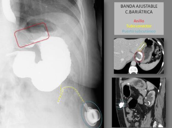
DISPOSITIVOS MEDICOS DE ABDOMEN Y PELVIS
En la práctica clínica y quirúrgica diaria se utilizan una gran variedad de dispositivos tanto para el tratamiento como seguimiento de multitud de patologías abdominopélvicas, pudiendo ser temporales o permantes, y constituyendo muchos de ellos hallazgos incidentales en los estudios radiológicos.
Además las técnicas de diagnóstico por imagen juegan un papel relevante en la determinación del correcto posicionamiento y función de estos sistemas.
Las complicaciones generales asociadas al uso de los mismos son migración, malposición y rotura, que pueden ser diagnosticados con rx simple; así como hemorragia, infección y lesión visceral, detectables con TC.
Descargas
Citas
Kim JS, Kim HC, Kim SW, Yang DM, Ryu JK, Rhee SJ, Kwon SH. Complications related to medical devices of the abdomen and pelvis: pictorial essay. Jpn J Radiol. 2015 Apr;33(4):177-86.
Gayer G, Lubner MG, Bhalla S, Pickhardt PJ. Imaging of abdominal and pelvic surgical and postprocedural foreign bodies. Radiol Clin North Am. 2014 Sep;52(5):991-1027.
Sonavane SK, Menias CO, Kantawala KP, Shanbhogue AK, Prasad SR, Eagon JC, Sandrasegaran K. Laparoscopic adjustable gastric banding: what radiologists need to know. Radiographics. 2012 Jul-Aug;32(4):1161-78.
Mausner EV, Yitta S, Slywotzky CM, Bennett GL. Commonly encountered foreign bodies and devices in the female pelvis: MDCT appearances. AJR Am J Roentgenol. 2011 Apr;196(4):W461-70.
Bahrami S, Chow D, Kadell B. AJR Am J Roentgenol. Thoracic and abdominal devices radiologists should recognize: pictorial review. 2009 Dec;193(6 Suppl):S106-18.
Smith HS, Deer TR, Staats PS, Singh V, Sehgal N, Cordner H. Intrathecal drug delivery. Pain Physician. 2008 Mar;11(2 Suppl):S89-S104.
Taljanovic MS, Hunter TB, Freundlich IM, Mar WA, Smyth SH, O'Brien MJ. Misplaced devices in the chest, abdomen, and pelvis: Part I. Semin Ultrasound CT MR. 2006 Apr;27(2):78-97.
Taljanovic MS, Hunter TB, Freundlich IM, Mar WA, Smyth SH, O'Brien MJ. Misplaced devices in the chest, abdomen, and pelvis: Part II. Semin Ultrasound CT MR. 2006 Apr;27(2):98-110.
Taljanovic MS, Hunter TB, O'Brien MJ, Schwartz SA. Gallery of medical devices: part 2: devices of the head, neck, spine, chest, and abdomen. Radiographics. 2005 Jul-Aug;25(4):1119-32.
Hunter TB, Taljanovic MS. Medical devices of the abdomen and pelvis. Radiographics. 2005 Mar-Apr;25(2):503-23.
O'Connor AR, Coakley FV. Retained surgical materials in the postoperative abdomen and pelvis. Semin Ultrasound CT MR. 2004 Jun;25(3):290-302.