Diagnóstico diferencial del dolor en fosa ilíaca derecha en la mujer, más allá de la apendicitis
Palabras clave:
dolor en fosa ilíaca derecha, mujer, poster, seram, FIDResumen
Objetivos Docentes
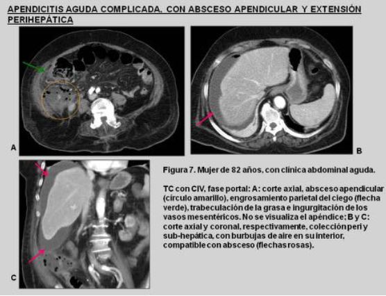
Ilustrar la anatomía normal de la fosa ilíaca derecha (FID) en la mujer sana y describir los diferentes hallazgos radiológicos de la patología aguda en FID, tanto con ecografía como con TC. (Figuras 1, 2 y 3).
Revisión del tema
La anatomía de la mujer a nivel de fosa ilíaca derecha es compleja y se ve influenciada por cambios hormonales y físicos, por lo que es importante conocer las diferentes entidades patológicas en función del grupo de edad. En este póster se revisan retrospectivamente estudios ecográficos y de TC de mujeres en edad fértil y postmenopáusicas, con dolor agudo en FID. Se ilustran los hallazgos radiológicos, dividiéndolos en patología intestinal, ginecológica y miscelánea mediante estas técnicas, exponiéndose los datos clave en la imagen para la realización de un adecuado diagnóstico diferencial. (Figura 4).
Descargas
Citas
: Purysko AS, Remer EM, Filho HM, Bittencourt LK, Lima RV, Racy DJ. Beyond appendicitis: common and uncommon gastrointestinal causes of right lower quadrant abdominal pain at multidetector CT. Radiographics. 2011 Jul-Aug;31(4):927-47.
: Kim YH, Blake MA, Harisinghani MG, Archer-Arroyo K, Hahn PF, Pitman MB,Mueller PR. Adult intestinal intussusception: CT appearances and identification of a causative lead point. Radiographics. 2006 May-Jun;26(3):733-44.
: Hyodoh H, Hori M, Akiba H, Tamakawa M, Hyodoh K, Hareyama M. Peripheral vascular malformations: imaging, treatment approaches, and therapeutic issues.Radiographics. 2005 Oct;25 Suppl 1:S159-71.
: Shadbolt CL, Heinze SB, Dietrich RB. Imaging of groin masses: inguinal anatomy and pathologic conditions revisited. Radiographics. 2001 Oct;21 Spec No:S261-71.
: Yitta S, Hecht EM, Slywotzky CM, Bennett GL. Added value of multiplanar reformation in the multidetector CT evaluation of the female pelvis: a pictorial review. Radiographics. 2009 Nov;29(7):1987-2003.
: Outwater EK, Siegelman ES, Hunt JL. Ovarian teratomas: tumor types and imaging characteristics. Radiographics. 2001 Mar-Apr;21(2):475-90.
: Chang HC, Bhatt S, Dogra VS. Pearls and pitfalls in diagnosis of ovarian torsion. Radiographics. 2008 Sep-Oct;28(5):1355-68.
: Jung SE, Lee JM, Rha SE, Byun JY, Jung JI, Hahn ST. CT and MR imaging of ovarian tumors with emphasis on differential diagnosis. Radiographics. 2002 Nov-Dec;22(6):1305-25.