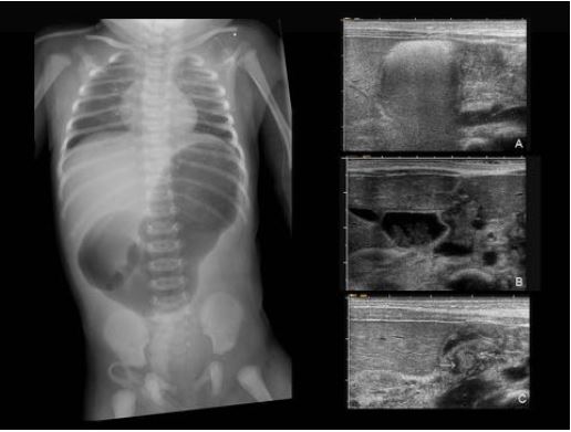
Distensión abdominal en el neonato:
más allá de la NEC...
Palabras clave:
Distensión abdominal, poster, seram, RN, neonatoResumen
Objetivos Docentes
La distensión del abdomen es el signo clínico más frecuente en la patología abdominal del recién nacido, siendo a su vez el principal motivo de las peticiones radiológicas. Los cuadros clínicos que se manifiestan como distensión abdominal entre otros signos y síntomas, así como su gravedad y pronóstico son muy diversos. Su adecuado y precoz diagnóstico es generalmente complejo, requiriendo un abordaje multidisciplinario (neonatólogos, cirujanos, radiólogos, anestesistas); fundamental dada la alta morbi-mortalidad de algunas patologías.
Revisión del tema
Índice de contenidos:
1. CONCEPTOS GENERALES.
2. INFORMACIÓN CLÍNICA Y PRUEBAS DE IMAGEN.
3. CAUSAS Y ENTIDADES QUE CURSAN CON DISTENSIÓN ABDOMINAL:
A) DISTENSION DE ORIGEN INTESTINAL:
1. OBSTRUCCIÓN
- ALTA
- BAJA
2. PSEUDO-OBSTRUCCIÓN / ILEO PARALITICO
3. PERFORACIÓN – PERITONITIS.
Descargas
Citas
Stephanie Ryan & Veronica Donoghue, Gastrointestinal pathology in neonates: new imaging strategies, Pediatr Radiol (2010) 40:927–931.
Veyrac C. et al., US assessment of neonatal bowel (necrotizing enterocolitis excluded). Pediatr Radiol. 2012;42 (Suppl 1):S104-7.
Charles M. Maxfield et al., A pattern-based approach to bowel obstruction in the newborn, Pediatr Radiol (2013) 43:318–329.
Cicero T. Silva et al., A prospective comparison of intestinal sonography and abdominal radiographs in a neonatal intensive care unit, Pediatr Radiol (2013) 43:1453–1463.
Rao P. et al., Neonatal gastrointestinal imaging. Eur J Radiol. 2006;60:171-206.
M.N. de la Hunt et al., The acute abdomen in the newborn, Seminars in Fetal & Neonatal Medicine (2006) 11, 191e197.
Arnold C. Merrow, Case 1: a newborn with bilious emesis. Pediatr Radiol (2014) 44:1462–1469.
Harris L. Cohen et al., The Vomiting Neonate or Young Infant, Ultrasound Clin 5 (2010) 97–112.
Hwa-Young K. et al., Bowel sonography in sepsis with pathological correlation: An experimental study. Pediatr Radiol (2011) 41:237–243.
Corinne Veyrac et al., US assessment of neonatal bowel (necrotizing enterocolitis excluded). Pediatr Radiol (2012) 42 (Suppl 1):S107–S114.
Emily F. et al., Commonly Encountered Surgical Problems in the Fetus and Neonate. Pediatr Clin N Am 56 (2009) 647–669.
Berrocal T. et al., Congenital Anomalies of the small intestine, colon and rectum. Radiographics 1999; 19:1219-1236.
Berrocal T. et al., Congenital Anomalies of the upper gastro-intestinal tract. Radiographics 1999; 19:855-872.
T. Berrocal, G. del Pozo et al., Gastrointestinal Emergencies in the Neonate. Capítulo 1.1. del libro Radilogical Imaging of the Digestive Tract in Intants and Children. A.S. Devos, J.G. Blickman 2008. Springer.
Alecia W. S. et al., Diagnostic performance of the upper gastrointestinal series in the evaluation of children with clinically suspected malrotation. Pediatr Radiol (2008) 38:518–528.
E. Ballesteros Gómiza et al., Malrotación-vólvulo intestinal: hallazgos radiológicos, Radiología. 2015;57(1):9---21.
John Amodio et al., Microcolon of Prematurity: A Form of Functional Obstruction, AJR 146:239-244, February 1986.
Richard J. Leone et al., Spontaneous’ Neonatal Gastric Perforation: Is It Really Spontaneous? Journal of Pediatric Surgery, Vol 35, No 7 (July), 2000: pp 1066-1069.
Praveen M. et al., Role of plain abdominal radiographs in predicting type of congenital pouch colon. Pediatr Radiol (2010) 40:1603–1608.
Justyna Romanska-Kita et al., Congenital chylous ascites. Pol J Radiol, 2011; 76(3): 58-61.


