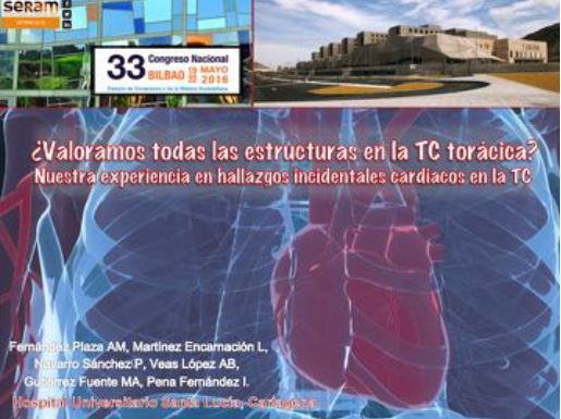
¿Valoramos todas las estructuras en la TC torácica?
Nuestra experiencia en hallazgos incidentales cardiacos en la TC.
Palabras clave:
hallazgos incidentales cardiacos, TC, poster, seram, angio-TC torácicaResumen
Objetivos Docentes
- Describir los hallazgos cardiacos más frecuentes que pueden hallarse incidentalmente en TC y angio-TC torácica y mostrar ejemplos de la casuística acontecida en nuestro Centro.
- Mostrar la importancia de analizar el corazón en la TC independientemente del motivo por el que se realice.
Revisión del tema
La TC es una de las técnicas de imagen más usadas en la valoración de la patología torácica, y el corazón suele pasar desapercibido. Sin embargo, no es infrecuente la presencia de
alteraciones inesperadas a nivel cardiaco. Algunas de estas serán variantes anatómicas, otras justificarán la clínica del paciente y otras no tendrán una correlación clínica clara, pero sí,
importantes consecuencias en el manejo de los pacientes. Por ello, es necesario incluir el corazón en la lectura sistemática de la TC torácica.
Descargas
Citas
- J. Kirsch, I. Buitrago, T.L. Mohammed, T. Gao, C.R. Asher, G.M. Novaro, Detection of coronary calcium during standard chest computed tomography correlates with multi-detector computed tomography coronary artery calcium score, Int. J. Cardiovasc. Imaging 28 (June (5)) (2012) 1249–1256.
- S.H. Lee, J.B. Seo, J.W. Kang, E.J. Chae, S.H. Park, T.H. Lim, Incidental cardiac and pericardial abnormalities on chest CT, J. Thorac. Imaging 23 (August (3)) (2008) 216–226.
- H.K. Kok, B. Loo, W.C. Torreggiani, O. Buckley, Incidental cardiac findings on thoracic imaging, Can. Assoc. Radiol. J. 64 (November (4)) (2013) 325–332.
- C. Hague, G. Andrews, B. Forster, MDCT of a malignant anomalous right coronary artery, AJR Am. J. Roentgenol. 182 (March (3)) (2004) 617–618.
- Luis Afonso, MD, FACC et al. Myocardial cleft, crypt, diverticulum, or aneurysm? Does it really matter?. Cardiol.32,8.2009.
- S. Pujadas y cols. Hendidura miocárdica: una alteración anatómica para tener en cuenta. Rev Esp Cardiol. 2009, 62 (7): 820-34.
- Minette MS, Sahn DJ. Ventricular septal defects. Circulation. 2006; 114(20): 2190-7.
- Erol C, Seker M. Coronary artery anomalies: the prevalence of origination, course, and termination anomalies of coronary arteries detected by 64-detector computed tomography coronary angiography. Journal of computer assisted tomography. 2011;35(5):618-24.
- Von Ziegler F, Pilla M, McMullan L, Panse P, Leber AW, Wilke N, et al. Visualization of anomalous origin and course of coronary arteries in 748 consecutive symptomatic patients by 64-slice computed tomography angiography. BMC cardiovascular disorders. 2009;9:54.
- Taylor AJ, Rogan KM, Virmani R. Sudden cardiac death associated with isolated congenital coronary artery anomalies. Journal of the American College of Cardiology. 1992;20(3):640-7.
- Opolski MP, Pregowski J, Kruk M, Witkowski A, Kwiecinska S, Lubienska E, et al. Prevalence and characteristics of coronary anomalies originating from the opposite sinus of Valsalva in 8,522 patients referred for coronary computed tomography angiography. The American journal of cardiology. 2013;111(9):1361-7.
- Pahlavan PS, Niroomand F. Coronary artery aneurysm: a review. Clinical cardiology. 2006;29(10):439-43.
- Al Attar N, Sablayrolles JL, Nataf P. Giant atherosclerotic aneurysm of the left anterior descending artery. The Journal of thoracic and cardiovascular surgery. 2003;126(3):888-90.
- Chen JJ, Manning MA, Frazier AA, Jeudy J, White CS. CT angiography of the cardiac valves: normal, diseased, and postoperative appearances. Radiographics. 2009;29(5):1393-412.
- A.H. Mahnken, G. Mu¨hlenbruch, M. Das, J.E. Wildberger, H.P. Ku¨hl, R.W. Gu¨nther, M. Kelm, R. Koos, MDCT detection of mitral valve calcification: prevalence and clinical relevance compared with echocardiography, AJR Am. J. Roentgenol. 188 (May (5)) (2007) 1264–1269.
- L.M. Boxt, M.J. Lipton, R.Y. Kwong, F. Rybicki, M.E. Clouse, Computed tomography for assessment of cardiac chambers, valves, myocardium and pericardium, Cardiol. Clin. 21 (November (4)) (2003) 561–585.
-M. Xie, Y. Li, T.O. Cheng, X. Wang, Q. Lu, L. He, M. Fu, Pseudoaneurysm of the mitral-aortic intervalvular fibrosa, Int. J. Cardiol. 166 (June (1)) (2013) 2–7.