Anatomía radiológica y estadificación de las neoplasias de hipofaringe
Palabras clave:
neoplasias de hipofaringe, poster, seramResumen
Objetivos Docentes
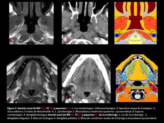
1) Describir la anatomía radiológica de la hipofaringe y su correlación con la anatomía macroscópica.
2) Revisar el estadiaje radiológico de las neoplasias de hipofaringe.
3) Identificar los cambios postratamiento de las neoplasias de hipofaringe.
Revisión del tema
1. ANATOMÍA DE LA FARINGE
EMBRIOLOGÍA
El sistema digestivo se desarrolla a partir del tubo intestinal primitivo, de origen endodérmico. La faringe deriva, embriológicamente, del intestino faríngeo que corresponde a la porción más cefálica del intestino anterior, y se extiende desde el estomodeo (o cavidad bucal primitiva) hasta el esbozo laringotraqueal.
Descargas
Citas
Marur S, Forastiere AA.Head and Neck Squamous Cell Carcinoma: Update on Epidemiology, Diagnosis, and Treatment.Mayo Clin Proc. 2016 Mar;91(3):386-96.
Paidpally V, Chirindel A, Lam S, Agrawal N, Quon H, Subramaniam RM.FDG-PET/CT imaging biomarkers in head and neck squamous cell carcinoma.Imaging Med. 2012 Dec;4(6):633-647.
Suenaga Y, Kitajima K, Kanda T, Otsuki N, Nibu KI, Sasaki R, Itoh T, Sugimura K.18F]-FDG PET/CT imaging for detection of nodal metastases in patients with squamous cell carcinoma
of the pharynx and larynx: comparison with CT.Jpn J Radiol. 2015 Dec 15
Silverman PM, Bossen EH, Fisher SR, Cole TB, Korobkin M, Halvorsen RA.Carcinoma of the larynx and hypopharynx: computed tomographic-histopathologic correlations. Radiology.
Jun;151(3):697-702.
American Joint Committee on Cancer AJCC cancer staging manual, 7TH ed Springer Science and Business Media LLC (SBM); 2010
Becker M, Zbären P, Delavelle J, Kurst AM, Egger C, Rufenacht A, Terrier F Neoplastic invasion of the laryngeal cartilage: reassessment of criteria for diagnosis at CT. Radiology. 1997.
:521–532
Xing Y, Zhang J, Lin H, Gold KA, Sturgis EM, Garden AS, Lee JJ, William WN Jr.Relation between the level of lymph node metastasis and survival in locally advanced head and neck
squamous cell carcinoma.Cancer. 2016 Feb 15;122(4):534-45.
Saito N, Nadgir RN, Nakahira M, Takahashi M, Uchino A, Kimura F, Truong MT, Sakai O.Posttreatment CT and MR imaging in head and neck cancer: what the radiologist needs to know. Radiographics. 2012 Sep-Oct;32(5):1261-82; discussion 1282-4.
Quon H, Brizel DM.Predictive and prognostic role of functional imaging of head and neck squamous cell carcinomas.Semin Radiat Oncol. 2012 Jul;22(3):220-32