“Anatomía y patología venosa intrahepática. Lo que el radiólogo debería conocer.”
Palabras clave:
patología venosa intrahepática, anatomía venosa intrahepática, poster, seramResumen
Objetivos Docentes
Repasar la anatomía vascular del hígado y revisar la patología venosa (portal y suprahepática) más frecuente.
Revisión del tema
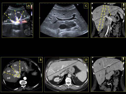
1. ANATOMÍA VENOSA HEPÁTICA
El hígado recibe un doble aporte vascular de la arteria hepática (15-25 %) y la vena porta (75-85 %) a través del hilio (porta hepatis). El drenaje venoso se realiza mediante las venas suprahepáticas a la cava inferior (VCI), que desemboca en la aurícula derecha. La vascularización venosa portal y sistémica no solo es importante para el correcto funcionamiento hepático, sino también para dividir el órgano en unidades (lóbulos y segmentos) que pueden tratarse de forma independiente.
Descargas
Citas
Withers CE, et al. Capítulo 4. Hígado. Rumack CM, et al. Diagnóstico por ecografía. Ed. Marbán, Madrid, 1999. pp 87-154.
Heiken JP, et al. Capítulo 12. Hígado. Lee JKT, et al. Body TC con correlación RM. Ed. Marbán, Madrid, 2007. pp 829-930.
Zwiebel WJ. Capítulo 28. Anatomía y características normales de la ecografía Doppler de los vasos del abdomen. Ziwiebel`s Doppler General. Zwiebel WJ, et al. Ed. Marbán, Madrid, 2008. pp 455-68.
Zwiebel WJ. Capítulo 32. Ecografía de la vasculatura hepática. Ziwiebel`s Doppler General. Zwiebel WJ, et al. Ed. Marbán, Madrid, 2008. pp 517-37.
Alustiza Echevarría JM y Martí-Bonmatí L. Capítulo 37. Radiología de las enfermedades hepáticas: enfermedad hepática difusa y lesión focal hepática. Del Cura JL, et al. Radiología
esencial. Ed. Panamericana, Madrid 2010. pp 512-25.
Covey AM, et al. Incidence, patterns, and clinical relevance of variant portal vein anatomy. AJR 2004; 183:1055-64.
Gallego C, et al. Congenital and acquiered anomalies of the portal venosus system. Radiographics 2002; 22:141-159.
Tirumani SH, et al. Imaging of the porta hepatis: spectrum of disease. Radiographics 2014; 34:73-92.
Torabi M, et al. CT of nonneoplastic hepatic vascula and perfusion disorders. Radiographics 2008; 28: 1967-82.
Katsuyoshi I, et al. CT of acquired abnormalities of the portal venosus system. Radiographics 1997; 17:897-917.
Tandon R, et al. Pylephlebitis after CT-guided percutaneous liver biopsy. AJR 2005; 184:70-72.
Farah M, et al. Ultrasound Case of the day. Radiographics 1992; 12:839-42.
Bhargava P, et al. Hepatic vascular shunts: embryology and imaging appearances. The British Journal of Radiology 2011; 84:1142-52.
Gallego C, ett al. Congenital hepatic shunts. Radigraphics 2004; 24:755-72.
Sfyroeras, GS, et al. Visceral venous aneurysms:clinical presentation, natural history and their management: a systematic review. Eur J Vasc Endovasc Surg 2009; 38:498-505.