PET/TC EN EL DIAGNÓSTICO, ESTADIAJE Y SEGUIMIENTO DE LOS LINFOMAS EN PEDIATRÍA
Palabras clave:
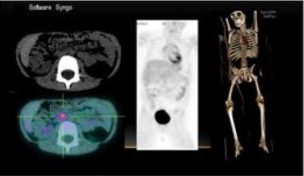
LINFOMAS, poster, seram, PEDIATRÍAResumen
Objetivos Docentes
- Exponer los principios técnicos de la PET/TC desde la preparación del paciente, la irradiación que implica la prueba, y la posibilidad de la utilización del contraste i.v. en base a la
importancia de una buena comunicación multidisciplinar.
- Conocer las indicaciones oncológicas de la PET/TC para el diagnóstico, seguimiento y estadiaje de los linfomas en pediatría, asi como los hallazgos que permiten establecer la
progresión, estabilidad o remisión de la enfermedad con esta prueba.
- Reconocer los potenciales pitfalls en la interpretación de las imágenes de la PET/TC, así como sus limitaciones.
Revisión del tema
INTRODUCCIÓN
Las complejas decisiones médicas sobre el tratamiento de los pacientes oncológicos son mayormente guiadas por los hallazgos en la imagen, entre otros factores. Muchos procedimientos radiológicos nos aportan información anatómica y morfológica de los tumores, pero poca o nula sobre su metabolismo. La tomografía computarizada (TC) y la resonancia magnética (RM) son técnicas de imagen que nos aportan información anatómica en el diagnóstico, estadiaje y seguimiento del cáncer.
Descargas
Citas
Sattar T, Griffeth LK, Latifi HR, Glass J, Munker R, Lilien DL. PET imaging today: contribution to the initial staging and prognosis of patients with non-Hodgkin’s lymphomas. J La State Med Soc 2006; 158(4):193–201.
Rini JN, Leonidas JC, Tomas MB, Palestro CJ. 18F-FDG PET versus CT for evaluating the spleen during initial staging of lymphoma. J Nucl Med 2003;44(7):1072–1074.
A.M. Álvarez Páez et al / Rev Esp Med Nucl Imagen Mol. 2012;31(6):340–349.
Mikhaeel NG, Hutchings M, Fields PA, O’Doherty MJ, Timothy AR. FDG-PET after two to three cycles of chemotherapy predicts progression-free and overall survival in high-grade non-Hodgkin lymphoma. Ann Oncol 2005;16(9):1514–1523.
Karam M, Novak L, Cyriac J, Ali A, Nazeer T, Nugent F. Role of fluorine-18 fluoro-deoxyglucose positron emission tomography scan in the evaluation and follow-up of patients with low-grade lymphomas. Cancer 2006;107(1):175–183.
Kazama T, Faria SC, Varavithya V, Phongkitkarun S, Ito H, Macapinlac HA. FDG PET in the evaluation of treatment for lymphoma: clinical usefulness and pitfalls. RadioGraphics
;25(1):191–207.
MR, Burger C, von Schulthess GK. PET/CT image co-registration in the thorax: influence of respiration. Eur J Nucl Med Mol Imaging 2002; 29: 351–360.