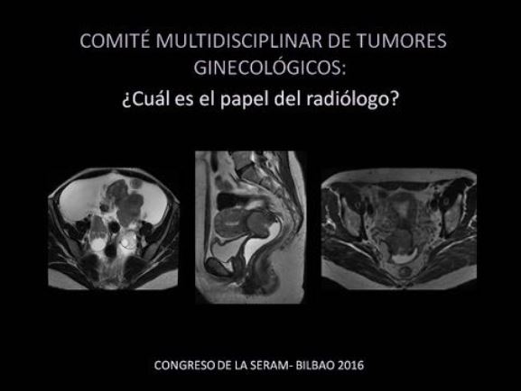
Comité Multidisciplinar de Tumores Ginecológicos:
¿cuál es el papel del radiólogo?
Palabras clave:
Comité Multidisciplinar de Tumores Ginecológicos, poster, seramResumen
Objetivos Docentes
Los comités multidisciplinares desempeñan un papel fundamental en el manejo de los pacientes oncológicos. En este póster mediante un formato interactivo se pretende mostrar el papel que tiene el radiólogo tanto en el diagnóstico y estadificación, como en la detección de posibles complicaciones y en la evolución de estos pacientes.
OBJETIVOS:
• Mostrar distintos casos clínicos y las preguntas más frecuentes planteadas por los distintos especialistas.
• Determinar las competencias del radiólogo en el diagnóstico, estadificación y monitorización en la respuesta al tratamiento/evolución.
• Exponer las diferentes técnicas de imagen empleadas, en especial la RM y el PET-TC.
Revisión del tema
Las pruebas de imagen juegan un papel fundamental en el estadiaje y valoración del grado de extensión de los distintos tumores ginecológicos y pueden ayudar al correcto manejo y tratamiento de estos pacientes.
En este póster mediante un formato interactivo se explican y muestran distintos casos presentados en el Comité Multidisciplinar de Tumores Ginecológicos, con las diferentes preguntas planteadas por los distintos especialistas (cirujanos, oncólogos, radioterapeutas) para un correcto manejo de estos pacientes.
Descargas
Citas
• Kyriazi S, Collins DJ, Morgan VA et al. Diffusion-weighted Imaging of peritoneal disease for Noninvasive Staging of Advanced Ovarian Cancer. Radiographics2010;30:1269-1285
• The Added Role of MR Imaging in treatment stratification of patients with Gynecologic Malignancies: What the radiologist needs to Know. Radiology 2013; 266(3): 717-740
• Rauch GM, Kaur H, Choi H et al. Optimization of MR Imaging for pretreatment Evaluation of patients with Endometrial and Cervical Cancer. Radiographics 2014; 34:1082-1098
• Dhanda S, Thakur M, Kerkar R, Jagmohan P. Diffusion-weight Imaging of Gynecologic Tumors: Diagnostic Pearls and Potencial Pitfalls. Radiographics 2014;34:1393-1416
• Chen J, Zhang Y, Liang B, Yang Z: The utility of diffusion-weighted MR imaging in cervical cancer. Eur. J. Radiol.2010;74:101-6
• Magne N, Chargari C, Vicenzi L et al. New trends in the evaluation and treatment of cervix cancer: the role of FDG-PET. Cancer Treat. Rev. 2008; 34: 671–81
• McVeigh PZ, Syed AM, Milosevic M, Fyles A,Haider MA. Diffusion-weighted MRI in cervical cancer. Eur Radiol 2008; 18:1058–64
• Chen J, Zhang Y, Liang B, Yang Z: The utility of diffusion-weighted MR imaging in cervical cancer. Eur. J. Radiol.2010;74:101-6
• Unger JB, Ivy JJ, Connor P, et al. Detection of recurrent cervical cancer by whole-body FDG PET scan in asymptomatic and symptomatic women. Gynecol Oncol 2004;94:212–6
• Grigsby PW, Siegel BA, Dehdashti F,Rader J, Zoberi I: Posttherapy (18F) fluorodeoxyglucose positron emission tomography in carcinoma of the cervix: response and outcome. J. Clin. Oncol. 2004 ;22: 2167–71
• Lin G, Ng KK, Chang CJ, Wang JJ, Ho KC, Yen TC, Wu TI, Wang CC, Chen YR, Huang YT, Ng SH, Jung SM, Chang TC, Lai CH. Myometrial invasion in endometrial cancer: diagnostic accuracy
of diffusion-weighted 3.0-T MR imaging--initial experience. Radiology. 2009;250:784-92
• Grigsby PW. Role of PET in gynecologic malignancy. Curr Opin Oncol. 2009; 21:420-4
• Whittaker CS, Coady A, Culver L, Rustin G, Padwick M, Padhani AR. Diffusion-weighted MR Imaging of Female Pelvic Tumors: A Pictorial Review. Radiographics 2009; 29:759-74
• Harry VN. Novel imaging techniques as response biomarkers in cervical cancer. Gynecol Oncol. 2010; 116:253-61
• Dilks P, Narayanan P, Reznek R, Sahdev A, Rockall A. Can quantitative dynamic contrast-enhanced MRI independently characterize an ovarian mass? Eur Radiol. 2010; 20 :2176-83
• Tempany CM, Zou KH, Silverman SG, Brown DL, Kurtz AB, McNeil BJ Staging of advanced ovarian cancer: comparison of imaging modalities-report from the Radiological Diagnostic
Oncology Group. Radiology 2000; 215 (3):761–767
• Fujii S, Kakite S, Nishihara K, Kanasaki Y, Harada T, Kigawa J, Kaminou T, Ogawa T. Diagnostic Accuracy of Diffusion-Weighted Imaging in Differentiating Benign From Malignant
Ovarian Lesions. J Magn Reson Imaging. 2008 ;28:1149-56
• Fujii S, Matsuse E, Kanasaki Y, et al. Detection of peritoneal dissemination in gynaecological malignancy: evaluation by diffusion-weighted MR imaging. Eur Radiol 2008; 18(1) 18–23
• Ungar L, Palfalvi L, Novak Z. Primary pelvic exenteration in cervical cancer patients. Gynecol Oncol 2008; 111:129
• Schneider A, Köhler C, Erdemoglu E. Current developments for pelvic exenteration in gynecologic oncology. Curr Opin Obstet Gynecol 2009; 21:4.
• Qayyum A, Coakley FV, Westphalen AC, et al. Role of CT and MR imaging in predicting optimal cytoreduction of newly diagnosed primary epithelial ovarian cancer. Gynecol Oncol 2005;96:301–6
• Sahdev A, Jones J, Shepherd JH, Reznek RH. MR imaging appearances of the female pelvis after trachelectomy. Radiographics. 2005 Jan-Feb;25(1):41-52


