Diagnóstico de las lesiones del ángulo pontocerebeloso
Palabras clave:
lesiones del ángulo pontocerebeloso, poster, seramResumen
Objetivos Docentes
• Conocer la anatomía del espacio pontocerebeloso para realizar una correcta valoración de las diferentes patologías.
• Describir los hallazgos radiológicos de las diferentes lesiones, las características y elementos clave del diagnóstico.
Revisión del tema
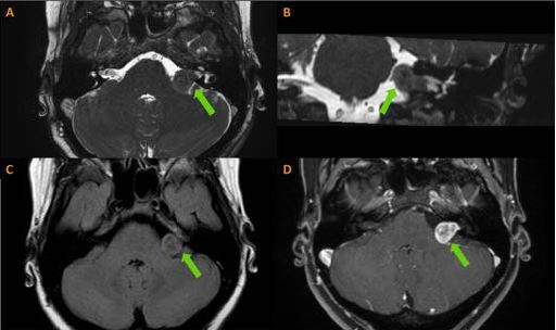
El área del ángulo pontocerebeloso (APC) es un espacio de pequeñas dimensiones 25 x 15 x 15 mm en la fosa posterior de la cavidad craneal. Dicho espacio está limitado anteriormente por la superficie posterior de la porción petrosa del hueso temporal y posteriormente por los hemisferios cerebelosos y superiormente por la tienda del cerebelo. El límite medial está formado por el núcleo olivar inferior y la cisterna de la protuberancia junto con el pedúnculo del cerebelo. El núcleo amigdalino del cerebelo forma el límite inferior de este espacio.
En el surco bulboprotuberancial emergen el VII y VIII pares craneales y se dirigen superior y lateral hacia el conducto auditivo interno. Cranealmente a estos pares se localiza el V par y caudalmente los nervios que
emergen del agujero rasgado posterior: IX, X y XI. Otras estructuras importantes en este espacio son los flóculos del cerebelo, la abertura lateral del cuarto ventrículo (foramen de Luschka) y la arteria cerebelosa
anteroinferior (AICA). La arteria laberíntica normalmente es una rama de la arteria cerebelosa anteroinferior, se divide en el conducto auditivo interno en tres ramas y proporciona irrigación a la cóclea, al laberinto anterior, a los nervios VIII en su porción coclear y al nervio facial en su porción endomeatal y laberíntica.
Descargas
Citas
• Juliano AF, Ginat DT, Moonis G. Imaging Review of the Temporal Bone: Part I. Anatomy and Inflammatory and Neoplastic Processes. Radiology, Oct 2013, Vol. 269: 17–33, 10.1148.
• Phillips GS, MD, LoGerfo SE, Richardson ML, et al. Interactive Webbased Learning Module on CT of the Temporal Bone: Anatomy and Pathology. RadioGraphics 2012; 32:E85–E105.
• Abdel Razek A, Huang BY. Lesions of the Petrous Apex: Classification and Findings at CT and MR Imaging. RadioGraphics 2012; 32:151–173.
• Osborn A, Salzman K, Barkovich J. Diagnostic imaging. Brain. 2011.
• Smirniotopoulos JG, Yue NC, Rushing EJ. Cerebellopontine angle masses: radiologicpathologic correlation. RadioGraphics 1993, Vol. 13: 1131–1147.
• Bonneville F, Sarrazin JC, MarsotDupuch K, et al Unusual Lesions of the Cerebellopontine Angle: A Segmental Approach. RadioGraphics 2001; 21:419–438.• Juliano AF, Ginat DT, Moonis G. Imaging Review of the Temporal Bone: Part I. Anatomy and Inflammatory and Neoplastic Processes. Radiology, Oct 2013, Vol. 269: 17–33, 10.1148.
• Phillips GS, MD, LoGerfo SE, Richardson ML, et al. Interactive Webbased Learning Module on CT of the Temporal Bone: Anatomy and Pathology. RadioGraphics 2012; 32:E85–E105.
• Abdel Razek A, Huang BY. Lesions of the Petrous Apex: Classification and Findings at CT and MR Imaging. RadioGraphics 2012; 32:151–173.
• Osborn A, Salzman K, Barkovich J. Diagnostic imaging. Brain. 2011.
• Smirniotopoulos JG, Yue NC, Rushing EJ. Cerebellopontine angle masses: radiologicpathologic correlation. RadioGraphics 1993, Vol. 13: 1131–1147.
• Bonneville F, Sarrazin JC, MarsotDupuch K, et al Unusual Lesions of the Cerebellopontine Angle: A Segmental Approach. RadioGraphics 2001; 21:419–438.


