DESCRIPCIÓN DE LOS DISPOSITIVOS INTRACRANEALES MÁS UTILIZADOS DESDE UN PUNTO DE VISTA RADIOLÓGICO.
Palabras clave:
DISPOSITIVOS INTRACRANEALES, poster, seramResumen
Objetivos Docentes
Los dispositivos intracraneales son herramientas que tienen como función la prevención, el diagnóstico, incluso la monitorización y el tratamiento de una gran variedad de patología cerebral.
El objetivo de este trabajo es revisar cada uno de los tipos de estos dispositivos así como su utilidad, sus componentes, su posición y sus posibles complicaciones.
Estos dispositivos son visualizados frecuentemente en estudios radiológicos. Es esencial para los radiológos conocer los distintos tipos que existen y su aplicación para así llevar a cabo una correcta
interpretación de los hallazgos de la imagen.
Revisión del tema
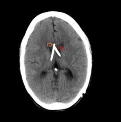
1. CATÉTERES DE DERIVACIÓN VENTRICULAR
1.1 HIDROCEFALIA
La Hidrocefalia (HCF) se define como un disbalance de la formación, circulación y absorción de liquido cefalorraquídeo, conduciendo a un incremento del volumen de este fluido en el SNC. Suelen asociarse a
HTIC (Hipertensión intracraneal).
Descargas
Citas
Tanaka N, Yamaguchi S, Ishikawa H, Ishii H, Meguro K. Prevalence of possible idiopathic normal-pressure hydrocephalus in Japan: the Osaki-Tajiri project. Neuroepidemiology. 2009. 32(3):171-5.
Klimo P Jr, Van Poppel M, Thompson CJ, Baird LC, Duhaime AC, Flannery AM, et al. Pediatric hydrocephalus: systematic literature review and evidence-based guidelines. Part 6: Preoperative antibiotics for shunt surgery in children with hydrocephalus: a systematic review and meta-analysis. J Neurosurg Pediatr. 2014 Nov. 14 Suppl 1:44-52
Whitehead WE, Riva-Cambrin J, Wellons JC 3rd, Kulkarni AV, Holubkov R, Illner A, et al. No significant improvement in the rate of accurate ventricular catheter location using ultrasound-guided CSF shunt insertion: a prospective, controlled study by the Hydrocephalus Clinical Research Network. J Neurosurg Pediatr. 2013 Oct 11.
Kazui H, Miyajima M, Mori E, Ishikawa M, SINPHONI-2 Investigators. Lumboperitoneal shunt surgery for idiopathic normal pressure hydrocephalus (SINPHONI-2): an open- label randomised trial. Lancet Neurol. 2015 Jun. 14 (6):585-94.
Manejo de los drenajes intraventriculares. Gobierno de Aragon. Sector Zaragoza. Protocolo de manejo UCI. Mayo 2012.
Izumihara A, Yamashita K, Murakami T. Acute subdural hematoma requiring surgery in the subacute or chronic stage. Neurol Med Chir (Tokyo). 2013. 53(5):323-8.
Tosaka M, Sakamoto K, Watanabe S, Yodonawa M, Kunimine H, Aishima K, et al. Critical classification of craniostomy for chronic subdural hematoma; safer technique for hematoma aspiration. Neurol Med Chir (Tokyo). 2013. 53(4):273-8.
[Guideline] Bullock MR, Chesnut R, Ghajar J, Gordon D, Hartl R, Newell DW, et al. Surgical management of acute subdural hematomas. Neurosurgery. 2006 Mar. 58(3 Suppl):S16-24; discussion Si-iv.
Muzii VF, Bistazzoni S, Zalaffi A, Carangelo B, Mariottini A, Palma L. Chronic subdural hematoma: comparison of two surgical techniques. Preliminary results of a prospective randomized study. J Neurosurg Sci. 2005 Jun. 49(2):41-6; discussion 46-7.
Li H, Pan R, Wang H, Rong X, Yin Z, Milgrom DP, et al. Clipping versus coiling for ruptured intracranial aneurysms: a systematic review and meta-analysis. Stroke. 2013 Jan. 44(1):29-37.
van Rooij WJ, Sprengers ME, de Gast AN, Peluso JP, Sluzewski M. 3D rotational angiography: the new gold standard in the detection of additional intracranial aneurysms. AJNR Am J Neuroradiol. 2008 May. 29(5):976-9.