EL MAPA NEURAL:
UNA NUEVA HERRAMIENTA PARA LA VALORACIÓN DE LAS POLINEUROPATÍAS
Palabras clave:
MAPA NEURAL, poster, seram, POLINEUROPATÍAS, enfermedades del nervio periférico, NPResumen
Objetivos Docentes
El propósito de este trabajo es contestar a estas cinco preguntas:
1.-¿Por qué estudiar ecográficamente las polineuropatías (PN)?
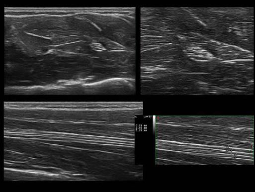
2.- ¿Qué es el mapa neural ecográfico?
3.- ¿Qué sabemos, hoy, de la ecografía en las PN?
4.- ¿Cuál es la aplicación del mapa neural, hoy, en la práctica clínica diaria?
5.- ¿Cuál es el futuro del mapa neural en el estudio de las PN?
Revisión del tema
1.-¿Por qué estudiar ecográficamente las polineuropatías (PN)?
Las enfermedades del nervio periférico (NP) son frecuentes en la práctica clínica, su diagnóstico se ha basado fundamentalmente en tres pilares: la historia clínica y exploración física, los estudios electrofisiológicos y pruebas de laboratorio; todos ellos dirigidos fundamentalmente a valorar la función.
Descargas
Citas
Beekman R, Visser LH. Sonographic detection of diffuse peripheral nerve enlargement in hereditary neuropathy with liability to pressure palsies. J Clin Ultrasound 2002;30(7):433-6.
Cartwright MS, Passmore LV, Yoon JS, Brown ME, Caress JB, Walker FO. Cross-sectional area reference values for nerve ultrasonography. Muscle & nerve 2008;37(5):566-71.
Fornage BD. Peripheral nerves of the extremities: imaging with US. Radiology 1988;167(1):179-82.
Gallardo E, Noto Y, Simon NG Ultrasound in the diagnosis of peripheral neuropathy: structure meets function in the neuromuscular clinic. J Neurol Neurosurg Psychiatry. 2015.Oct;86(10):1066-74. doi: 10.1136/jnnp-2014-309599. Epub 2015 Feb 4.
Gallardo E, Sedano MJ, Orizaola P, et al. Spinal nerve involvement in early Guillain-Barre syndrome: A clinico-electrophysiological, ultrasonographic and pathological study. Clinical
neurophysiology : official journal of the International Federation of Clinical Neurophysiology 2014 doi: 10.1016/j.clinph.2014.06.051[published Online First: Epub Date]|.
Ginanneschi F, Filippou G, Giannini F, et al. Sonographic and electrodiagnostic features of hereditary neuropathy with liability to pressure palsies. Journal of the peripheral nervous system : JPNS 2012;17(4):391-8.
Goedee HS, Brekelmans GJF, van Asseldonk JTH, Beekman R, Mess WH. High resolution sonography in the evaluation of the peripheral nervous system in polyneuropathy - a review of the literature. European journal of neurology 2013;20:1342-51.
Goedee HS, Brekelmans GJ, Visser LH. Multifocal enlargement and increased vascularization of peripheral nerves detected by sonography in CIDP: a pilot study. Clinical neurophysiology
;125(1):154-9.
Haun DW, Cho JC, Kettner NW. Normative cross-sectional area of the C5-C8 nerve roots using ultrasonography. Ultrasound in medicine & biology 2010;36(9):1422-30.
Jain S, Visser LH, Praveen TL et al. High-resolution sonography: a new technique to detect nerve damage in leprosy. PloS neglected tropical disease 2009; 3: 498.
Kerasnoudis A, Pitarokoili K, Behrendt V, Gold R, Yoon MS. Correlation of Nerve Ultrasound, Electrophysiological and Clinical Findings in Chronic Inflammatory Demyelinating Polyneuropathy. Journal of neuroimaging : official journal of the American Society of Neuroimaging 2014 doi: 10.1111/jon.12079[published Online First: Epub Date]|.
Kerasnoudis A, Pitarokoili K, Behrendt V, Gold R, Yoon MS. Nerve ultrasound score in distinguishing chronic from acute inflammatory demyelinating polyneuropathy. Clinical neurophysiology : official journal of the International Federation of Clinical Neurophysiology 2014;125(3):635-41.
Martinoli C, Schenone A, Bianchi S, et al. Sonography of the median nerve in Charcot-Marie-Tooth disease. AJR. American journal of roentgenology 2002;178(6):1553-6.
Martinoli C, Derchi LE, Bertolotto M, et al. US and MR imaging of peripheral nerves in leprosy. Skeletal radiology 2000;29(3):142-50.
Noto YI, Shiga K, Tsuji Y, et al. Nerve ultrasound depicts peripheral nerve enlargement in patients with genetically distinct Charcot-Marie-Tooth disease. Journal of neurology, neurosurgery, and psychiatry 2014 doi: 10.1136/jnnp-2014-308211[published Online First: Epub Date]|.
Padua L, Martinoli C, Pazzaglia C, et al. Intra- and internerve cross-sectional area variability: new ultrasound measures. Muscle & nerve 2012;45(5):730-3.
Pazzaglia C, Minciotti I, Coraci D, Briani C, Padua L. Ultrasound assessment of sural nerve in Charcot-Marie-Tooth 1A neuropathy. Clinical neurophysiology : official journal of the International Federation of Clinical Neurophysiology 2013;124(8):1695-9