LA RESISTENCIA EN LA BATALLA TERAPEUTICA PERCUTÁNEA CONTRA LOS LINFANGIOMAS:
ANALISIS DEL NÚMERO DE SESIONES Y ÉXITO EN FUNCIÓN DEL ESCLEROSANTE EMPLEADO.
Palabras clave:
LINFANGIOMAS, poster, seram, malformaciones linfáticas, MLResumen
Objetivos
Las malformaciones linfáticas o linfangiomas (ML), se consideran malformaciones vasculares congénitas de bajo flujo según la clasificación de Mullicken y Glowacki (1982) [1], aunque actualmente existe un debate sobre su teórica capacidad proliferativa [2]. Consisten en una conexión anómala y aislada de la circulación general de los vasos linfáticos que provoca espacios quísticos rellenos de linfa.
En función del tamaño de sus quistes, se clasifican en macroquísticos (quistes > 2cm), mixtos y microquísticos (quistes < 2 cm en diámetro). Pueden tener localización superficial o profunda [2-4]. De forma frecuente asocian hipertrofia del tejido celular subcutáneo adyacente. La ecografía es la técnica diagnóstica inicial, aunque la resonancia magnética es la prueba de elección para su confirmación, permitiendo analizar la extensión de la lesión, su grado de combinación con otras malformaciones y complicaciones [5-7].
Material y métodos
Procedimiento y protocolo terapéutico
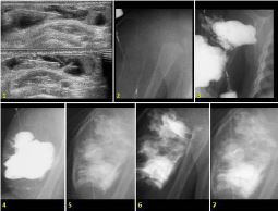
Los linfangiomas incluidos presentaban al menos una ecografía en nuestro centro previa a la intervención percutánea, de carácter diagnóstico y/o evaluación preoperatoria. Se empleó el modo B y modo Doppler color (Sequoia 512 Acuson Siemens), con transductor de 7,5 – 14 MHz, para valorar tipo de linfangioma, morfología, localización, extensión, relaciones anatómicas y vascularización. En casos de ecografías no concluyentes o sospecha de complicaciones, se realizó RM posterior con protocolo habitual (Figura 1).
Descargas
Citas
Mulliken JB, GlowackiJ. Hemangiomas and vascular malformations in infants and children: a classification based on endothelial characteristics. PlastReconstrSurg 1982; 69:412–420
Garzon M, Huang J, Enjolras O, Frieden I. Vascular malformations. Part I. J Am AcadDermatol 2007;56:353-70
Puig S, Casati B, Staudenherz A, Paya K. Vascular low-flow malformations in children: current concepts for classification, diagnosis and therapy. European Journal of Radiology 2005; 53: 35 – 45.
Burrows P, Mason K. Precutaneous treatment of low flow vascular malformations. J Vas IntervRadiol 2004; 15: 431 – 445.
Dobous J. Garel L. Imaging and therapeutic approach of hemangiomas and vascular malformations in the pediatric age group. PediatrRadiol 1999; 29: 879 – 893.
Shiels II W, Kenney B, Caniano D, Besner G. Definitive percutaneos treatment of lymphatic malformations of the trunk and extremities. Journal of Pediatric surgery 2008; 43: 136 – 140.
Dubois J, Alison M. Vascular malformations: what a radiologist needs to know. PediatrRadiol 2010; 40: 895 – 905.
Burrows P, Mitri R, Alomari A, Padua H, Lord D, Sylvia M, et al. Precutaneoussclerotherapy of lymphatic malformations with doxycicline. Lymphatic research and biology 2008; 6 (4).
Acevedo J, Shah R, Brietzke S. Nonsurgical therapies for lymphamgiomas: a systematic review. Otolaryngology – head and neck surgery 2008; 138: 418 – 424.
Giguere CM, Bauman NM, Smith RJ. New treatment options for lymphangioma in infants and children. Ann OtlRhinolLaryngil 2002; 111: 1006 – 75.
Alomari A, Karian V, Lord D, Padua H, Burrows P. Percutaneoyssclerotherapy for lymphatic malformations: a retrospective analysis of patient – evaluated improvement. J VascIntervRadiol 2006; 17: 1639 – 1648.
Yoo J, Ahn Y, Lim Y, Hah J, Kwon T, Sung M, et al. OK432 sclerotherapy in head and neck lymphangiomas: long-term follow up result. Otolaryngology – head and neck surgery 2009; 140: 120 – 123.
Jain R, Bandhu S, Sawhney S, Mittal R. Sonographically guided percutaneous sclerosis using 1% polidocanol in treatment of vascular malformations. Journal of clinical ultrasound 2002; 30 (7): 416 – 423.


