Imagen de difusión en Resonancia Magnética abdominopélvica:
Técnica y utilidad.
Palabras clave:
Resonancia Magnética abdominopélvica, poster, seramResumen
Objetivos Docentes
Revisar los aspectos físicos básicos de la técnica de difusión en Resonancia Magnética, asì como también las ventajas, utilidades, aplicaciones, limitaciones y potenciales errores en la patología abdominopélvica.
Revisión del tema
Introducción
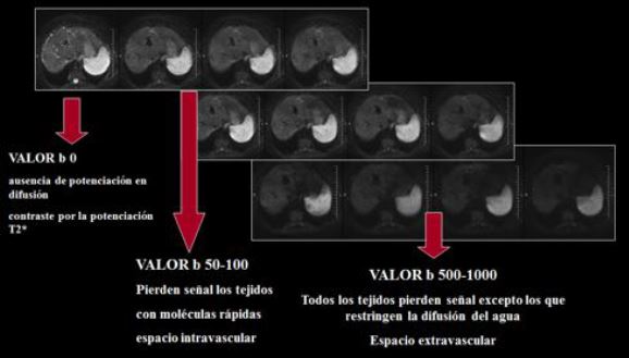
Desde los años 90, una serie de avances tecnológicos hicieron posible la aplicación de la difusión en RM a regiones extracraneales, incluyendo el abdomen y pelvis. El desarrollo de la imagen ecoplanar, gradientes de alta amplitud, bobinas multielemento y la adquisición en paralelo han sido imprescindibles.
La adquisición en paralelo, mediante la reducción del tiempo de eco (TE), la longitud de trenes de eco y el tiempo de llenado del espacio K, permite adquisiciones rápidas con menos artefactos de movimiento, obteniendo imágenes de difusión de alta calidad.
Descargas
Citas
Koh D-M, Blackledge M, Padhani AR, Takahara T, Kwee TC, Leach MO, et al. Whole-Body Diffusion-Weighted MRI: Tips, Tricks, and Pitfalls. American Journal of Roentgenology. 2012 Aug;199(2):252–62.
Padhani AR, Miles KA. Multiparametric imaging of tumor response to therapy. Radiology. 2010 Aug;256(2):348–64.
Padhani AR, Koh D-M, Collins DJ. Whole-body diffusion-weighted MR imaging in cancer: current status and research directions. Radiology. 2011 Dec;261(3):700–18.
Padhani AR, Gogbashian A. Bony metastases: assessing response to therapy with whole-body diffusion MRI. Cancer Imaging. 2011;11(1A):S129–54.
Littooij AS, Kwee TC, Barber I, Granata C, Vermoolen MA, Enríquez G, et al. Whole-body MRI for initial staging of paediatric lymphoma: prospective comparison to an FDG-PET/CT-based reference standard. Eur Radiol. 2014 Feb 23;24(5):1153–65.
Byun WM, Shin SO, Chang Y, Lee SJ, Finsterbusch J, Frahm J. Diffusion-weighted MR imaging of metastatic disease of the spine: assessment of response to therapy. AJNR Am J Neuroradiol. 2002 Jun;23(6):906–12.
Adams HJA, Kwee TC, Vermoolen MA, Keizer B, Klerk JMH, Adam JA, et al. Whole-body MRI for the detection of bone marrow involvement in lymphoma: prospective study in 116 patients and comparison with FDG-PET. Eur Radiol. 2013 Apr 17;23(8):2271–8.
Phd ACMMJM, Md BVSG, Md ALJ, Phd AJDRM. Diffusion MR Imaging for Monitoring Treatment Response. Neuroimaging Clinics of NA. 2011 Feb 1;21(1):153–78.
Reischauer C, Froehlich JM, Koh D-M, Graf N, Padevit C, John H, et al. Bone metastases from prostate cancer: assessing treatment response by using diffusion-weighted imaging and functional diffusion maps--initial observations. Radiology. 2010 Nov;257(2):523–31.
Michielsen K, Vergote I, beeck K, Amant F, Leunen K, Moerman P, et al. Whole-body MRI with diffusion-weighted sequence for staging of patients with suspected ovarian cancer: a clinical feasibility study in comparison to CT and FDG-PET/CT. Eur Radiol. 2013 Dec 11;24(4):889–901.
Mowatt G, Scotland G, Boachie C, Cruickshank M, Ford J, Fraser C, et al. The diagnostic accuracy and cost-effectiveness of magnetic resonance spectroscopy and enhanced magnetic resonance imaging techniques in aiding the localisation of prostate abnormalities for biopsy: a systematic review and economic evaluation. Health Technol Assess. 2013 May;17(20):1–281.
Espada M, Garcia-Flores JR, Jimenez M, Alvarez-Moreno E, Haro M, Gonzalez-Cortijo L, et al. Diffusion-weighted magnetic resonance imaging evaluation of intra-abdominal sites of implants to predict likelihood of suboptimal cytoreductive surgery in patients with ovarian carcinoma. Eur Radiol. 2013 Apr 19;23(9):2636–42.
Luboldt W, Kufer R, Blumstein N, Toussaint TL, Kluge A, Seemann MD, et al. Prostate Carcinoma: Diffusion-weighted Imaging as Potential Alternative to Conventional MR and 11C-Choline PET/CT for Detection of Bone Metastases. Radiology. 2008 Nov 14;249(3):1017–25.
Koh D-M, Collins DJ. Diffusion-Weighted MRI in the Body: Applications and Challenges in Oncology. American Journal of Roentgenology. 2007 Jun;188(6):1622–35.
Mosavi F, Johansson S, Sandberg DT, Turesson I, Sörensen J, Ahlström H. Whole-Body Diffusion-Weighted MRI Compared With 18F-NaF PET/CT for Detection of Bone Metastases in
Patients With High-Risk Prostate Carcinoma. American Journal of Roentgenology. 2012 Nov;199(5):1114–20.
Afaq A. Diffusion-weighted magnetic resonance imaging for tumour response assessment: why, when and how? Cancer Imaging. 2010;10(1A):S179–88.
Lecouvet FE, Larbi A, Pasoglou V, Omoumi P, Tombal B, Michoux N, et al. MRI for response assessment in metastatic bone disease. Eur Radiol. 2013 Mar 1;23(7):1986–97.
Li SP, Padhani AR. Tumor response assessments with diffusion and perfusion MRI. J Magn Reson Imaging. 2012 Mar 20;35(4):745–63.
Wu L-M, Gu H-Y, Zheng J, Xu X, Lin L-H, Deng X, et al. Diagnostic value of whole-body magnetic resonance imaging for bone metastases: a systematic review and meta-analysis. J Magn Reson Imaging. 2011 May 25;34(1):128–35.
Dow-Mu Koh, David J. Collins. Diffusion-Weighted MRI in the Body: Applications and Challenges in Oncology. AJR 2007; 188:1622–1635.
Aliya Qayyum, MBBS, MRCP, FRCR. Diffusion-weighted Imaging in the Abdomen and Pelvis: Concepts and Applications. RadioGraphics 2009; 29:1797–1810.
Dell P. Dunn, Karen S. Lee, Martin P. Smith, Koenraad J. Mortel. Nononcologic Applications of Diffusion-Weighted Imaging in the Gastrointestinal System
Dell P. Dunn, et al. Non-oncologic applications of diffusionweighted imaging (DWI) in the genitourinary system. Abdom Imaging (2015) 40:1645–1654
William A. Moore, Gaurav Khatri, et al. Added Value of Diffusion-Weighted Acquisitions in MRI of the Abdomen and Pelvis. AJR 2014; 202:995–1006.