PAPEL DE LA RM EN LOS PACIENTES CON DIABETES INSÍPIDA.
Palabras clave:
DIABETES INSÍPIDA, poster, seramResumen
Objetivos Docentes
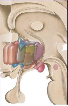
Realizaremos un repaso de la anatomía de la hipófisis.
Revisión del comportamiento en imagen de la neurohipófisis en los distintos procesos patológicos que pueden cursar con Diabetes Insípida.
Revisión del tema
INTRODUCCIÓN
La diabetes insípida (DI) es una patología que cursa con una alteración en la regulación del agua corporal, como consecuencia de una secreción o acción reducidas de la ADH (hormona antidiurética). La hormona antidiurética (ADH o vasopresina) es sintetizada a nivel de los núcleos supraóptico y paraventricular del hipotálamo y almacenada en la neurohipófisis para su liberación posterior al torrente sanguíneo.
Descargas
Citas
Ouyang T, Rothfus WE, Ng JM and Challinor SM: Imaging of the pituitary. Radiol Clin N Am (2011) 49: 549-571.
Fujisawa I, Asato R, Nishimura K et al. Anterior and posterior lobes of the pitutitary gland: assessment by 1.5T MR imaging. J Comput Assist Tom (1987) 11:214-220.
Colombo R, Berry I, Kucharczyk J et al. Posterior pituitary gland: appearance in normal MRI in normal and pathological states. Radiology (1987) 165:481-485.
Kurikawa H, Fujisawa I, Nakano Y et al. Posterior lobe of the pituitary gland: correlation between signal intensity on T1-weighted MR images and vasopressin concentration. Radiology (1998) 207:79-83.
Saleem SN, Said AHM and Lee DH. Lesions of the hypothalamus: MR imaging diagnostic features. RadioGraphics (2007) 27:1087-1108.
Turcu AF, Erickson BJ, Lin E et al. Pituitary stalk lesions: the Mayo Clinic experience. J Clin Endocrinol Metab (2013) 98:1812-1818.
Hamilton BE, Salzman KL and Osborn AG. Anatomic and pathologic spectrum of pituitary infundibulum lesions. AJR (2007) 188:W223-232.
D’Ambrosio N, Soohoo S, Warshall C et al. Craneofacial and intracranial manifestations of Langerhans cell histiocytosis: report of findings in 100 patients. AJR (2008) 191:589-597.