El papel del Radiólogo en la valoración de lesiones de Cabeza y Cuello en la Era de la Cirugía Robótica
Palabras clave:
lesiones de Cabeza, lesiones de cuello, poster, seram, técnica robótica transoral, TORS, Cirugía RobóticaResumen
Objetivos Docentes
- Conocer las peculiaridades de la técnica robótica transoral (TORS) en la esfera de la patología de Cabeza y Cuello.
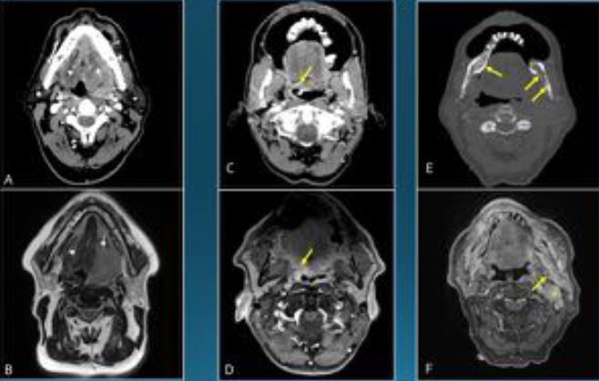
- Conocer el papel de las técnicas de imagen seccionales TC y RM en el manejo de pacientes candidatos a TORS con patología tanto maligna como benigna de Cabeza y Cuello.
Revisión del tema
INTRODUCCIÓN
La cirugía robótica fue concebida a principios de la pasada década inicialmente como un sistema de de teleasistencia en la que el potencial cirujano pudiera realizar una intervención estando físicamente alejado del paciente. Secundariamente se apreció su capacidad de optimizar los abordajes de cirugía de mínima invasión.
En la actualidad se trata de una técnica en expansión aunque con aplicaciones clínicas plenamente establecidas en diferentes especialidades como urología, ginecología, cirugía cardiotorácica y cirugía de Cabeza y Cuello.
El abordaje transoral mediante cirugía robótica - Transoral Robotic Surgery (TORS) - es una de las indicaciones consolidadas de esta técnica en Otorrinolaringología. Tiene como objetivo la exéresis de patología benigna y maligna de la vía aerodigestiva superior, fundamentalmente del cáncer de
orofaringe.
Descargas
Citas
2. Corey A. Pitfalls in the Staging of Cancer of the Oropharyngeal Squamous Cell Carcinoma. Neuroimaging Clin N Am. 2013;23(1):47–66. doi:10.1016/j.nic.2012.08.005.
3. Rovira Cañellas A., Siurana Monilva S., Tumores malignos de la naso-orfaringe y de la cavidad oral. In: De Juan Delgado M., Azpeitia Armán J. Actualizaciones SERAM Radiología de cabeza y cuello; 2012:44-58.
4. Tshering Vogel DW, Zbaeren P, Thoeny HC. Cancer of the oral cavity and oropharynx. Cancer Imaging. 2010;10:62–72. doi:10.1102/1470-7330.2010.0008.
5. Hudgins PA, Beitler JJ. I n t ro d u c t i o n t o t h e I m a g i n g and Staging of Cancer The tumor node metastasis staging. 2013;23:1–7.
6. Eisenmenger LB, Wiggins RH. Imaging of Head and Neck Lymph Nodes. Radiol Clin North Am. 2015;53(1):115–132. doi:10.1016/j.rcl.2014.09.011.
7. Cohan DM, Popat S, Kaplan SE, Rigual N, Loree T, Hicks WL. Oropharyngeal cancer: current understanding and management. Curr Opin Otolaryngol Head Neck Surg. 2009;17(2):88–94. doi:10.1097/MOO.0b013e32832984c0.
8. Hirunpat S, Jongsatitpaiboon J, Angunsri N, Chowchuvech V. When should MRI be recommended for the accurate clinical staging of base of tongue carcinoma? Asian Pacific J Cancer Prev. 2007;8(2):310–314.
9. Park J-O, Jung S-L, Joo Y-H, Jung C-K, Cho K-J, Kim M-S. Diagnostic accuracy of magnetic resonance imaging (MRI) in the assessment of tumor invasion depth in oral/oropharyngeal cancer. Oral Oncol. 2011;47(5):381–386. doi:10.1016/j.oraloncology.2011.03.012.
10. Jansen JF a, Schöder H, Lee NY, et al. Tumor metabolism and perfusion in head and neck squamous cell carcinoma: Pretreatment multimodality imaging with 1H magnetic resonance spectroscopy, dynamic contrast-enhanced MRI, and [ 18F]FDG-PET. Int J Radiat Oncol Biol Phys. 2012;82(1):299–307. doi:10.1016/j.ijrobp.2010.11.022.
11. Ng S-H, Lin C-Y, Chan S-C, et al. Clinical Utility of Multimodality Imaging with Dynamic Contrast-Enhanced MRI, Diffusion-Weighted MRI, and 18F-FDG PET/CT for the Prediction of Neck Control in Oropharyngeal or Hypopharyngeal Squamous Cell Carcinoma Treated with Chemoradiation.
12. Jansen JF1, Carlson DL, Lu Y, Stambuk HE, Moreira AL, Singh B, Patel SG, Kraus DH, Wong RJ, Shaha AR, Shah JP S-DA. Correlation of a priori DCE-MRI and (1)H-MRS data with molecular markers in neck nodal metastases: Initial analysis. Oral Oncol. 2012;48(6):717–22. doi:10.1016/j.oraloncology.2012.02.001.
13. Chawla S, Kim S. Pretreatment diffusion-weighted and dynamic contrast-enhanced MRI for prediction of local treatment response in squamous cell carcinomas of the head and neck. Am J …. 2013;200(1):35–43. doi:10.2214/AJR.12.9432.Pretreatment.
14. Ng SH, Lin CY, Chan SC, et al. Dynamic Contrast-Enhanced MR Imaging Predicts Local Control in Oropharyngeal or Hypopharyngeal Squamous Cell Carcinoma Treated with Chemoradiotherapy. PLoS One. 2013;8(8):1–11. doi:10.1371/journal.pone.0072230.
15. Preda L, Calloni SF, Moscatelli MEM, Cossu Rocca M, Bellomi M. Role of CT Perfusion in Monitoring and Prediction of Response to Therapy of Head and Neck Squamous Cell Carcinoma. Biomed Res Int. 2014;2014:917150. doi:10.1155/2014/917150.
16. Landry D. GC. Squamous cell carcinoma of the upper aerodigestive tract: a review. Radiol Clin North Am. 2015;53(1):81–97. doi:doi: 10.1016/j.rcl.2014.09.013.
17. Gamss C, Gupta A, Chazen JL, Phillips CD. Imaging Evaluation of the Suprahyoid Neck. Radiol Clin North Am. 2015;53(1):133–144. doi:10.1016/j.rcl.2014.09.009.
18. Trotta BM, Pease CS, Rasamny JJ, Raghavan P, Mukherjee S. Oral cavity and oropharyngeal squamous cell cancer: key imaging findings for staging and treatment planning. Radiographics. 2011;31(2):339–54. doi:10.1148/rg.312105107.
19. Sanders I, Mu L. A three-dimensional atlas of human tongue muscles. Anat Rec. 2013;296(7):1102–1114. doi:10.1002/ar.22711.
20. Lörincz BB, Jowett N, Knecht R. Decision management in transoral robotic surgery (TORS): indications, individual patient selection, and role in the multidisciplinary treatment of head and neck cancer from a european perspective. Head Neck. 2015;(Mar):Epub.
21. Hinni ML, Nagel T, Howard B. Oropharyngeal cancer treatment: the role of transoral surgery. Curr Opin Otolaryngol Head Neck Surg. 2015;23(2):132–138.
doi:10.1097/MOO.0000000000000143.
22. Weinstein GS, O’Malley BW Jr, Snyder W, Sherman E QH. Transoral robotic surgery: radical tonsillectomy. Arch Otolaryngol Head Neck Surg. 2007;133(12):1220–6. (6)
23. Loevner L a, Learned KO, Mohan S, et al. Transoral robotic surgery in head and neck cancer: what radiologists need to know about the cutting edge. Radiographics. 2013;33(6):1759–79. doi:10.1148/rg.336135518.
24. Arora A, Cunningham A, Chawdhary G, et al. Clinical applications of Telerobotic ENT-Head and Neck surgery. Int J Surg. 2011;9(4):277–284. doi:10.1016/j.ijsu.2011.01.008.
25. Hans S, Delas B, Gorphe P, Ménard M, Brasnu D. Transoral robotic surgery in head and neck cancer. Eur Ann Otorhinolaryngol Head Neck Dis. 2012;129(1):32–37. doi:10.1016/j.anorl.2011.06.003.
26. Rinaldi V, Pagani D, Torretta S, Pignataro L. Transoral robotic surgery in the management of head and neck tumours. Ecancermedicalscience. 2013;7(1):1–15. doi:10.3332/ecancer.2013.359.
27. Kucur C, Durmus K, Gun R, et al. Safety an efficacy of Concurrent neck dissection and transoral robotic surgery. Head Neck. 2015;Mar:Epub. doi:10.1002/acr.22212.
28. Granell J, Mendez-Benegassi I, Millas T, Garrido L, Gutierrez-Fonseca R. Transoral Robotic Surgery: Step-by-Step Radical Tonsillectomy. Case Rep Otolaryngol. 2014;2014:497528. doi:10.1155/2014/497528.