Mientras mirábamos la mama:
lesiones axilares incidentales
Palabras clave:
lesiones axilares incidentales, mama, poster, seramResumen
Objetivos Docentes
Revisar e ilustrar las lesiones axilares no linfáticas incidentales encontradas en estudios de mama con correlación histopatológica.
Evaluar la utilidad y limitaciones de la mamografía, ecografía y resonancia magnética en la valoración de estas lesiones.
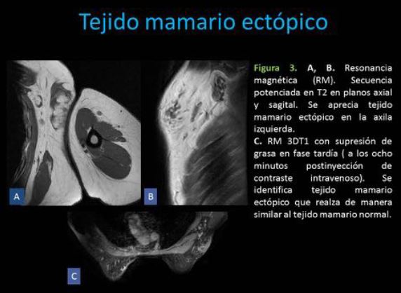
Revisión del tema
Anatomía
La axila es un espacio triangular entre el surco intertubercular del húmero lateralmente, el serrato anterior y pared torácica medialmente, pectoral mayor y menor y músculos subclavios anteriormente, y los músculos subescapular, redondo mayor y dorsal ancho posteriormente.
El ápex está delimitado por la primera costilla, la escápula y la clavícula. Fig. 1
Descargas
Citas
• Park Y.M., Park Ji-Sung, Yoon, H, Yang W. Imaging-Pathologic correlation of diseases in the axilla. AJR 2013; 200:W130-W142
• Leibman AJ, Wong R. Findings on mammography in the axilla. AJR 1997; 169: 1385-1390
• Kim EY, Ko EY, Han BK, Shin JH, Kang SS, Cho EY, Kim MJ, Chun SY. Sonography of axillary masses. J Ultrasound Med 2009; 28:923-939
• Giess CS, Raza S, Birdwell RL. Distinguishing breast skin lesions from superficial breast parenchymal lesions: diagnostic criteria, imaging characteristics, and pitfalls. Radiographics 2011; 31: 1959-1972.
• Filipovski V, Banev S, Janevska V, Dukova B. Granular cell tumor of the breast: a case report and review of literature. Cases Journal. 2009;2:8551. doi:10.4076/1757-1626-2-8551.
• Nasit JG, Chauhan S, Dhruva G. Granular cell tumor of hand presenting as subcutaneous nodule mimicking dermal adnexal tumor: A diagnosis by cytology. Indian Dermatology Online Journal. 2013;4(1):33-36. doi:10.4103/2229-5178.105467.
• Aoyama K, Kamio T, Hirano A, Seshimo A, Kameoka S. Granular cell tumors: a report of six cases. World J Surg Oncol 2012 Sep 29;10:204. doi: 10.1186/1477-7819-10-204.
• Dialani V, James DF, Slanetz PJ. A practical approach to imaging the axilla.Insights into Imaging. 2015;6(2):217-229. doi:10.1007/s13244-014-0367-8.
• Shir-Hwa Ueng, Thomas Mezzetti, and Fattaneh A. Tavassoli. Papillary Neoplasms of the Breast: A Review. Archives of Pathology & Laboratory Medicine 2009 133:6, 893-907 .
• Levy S, Samuels T, Catzavelos C, Hamilton P, Shumak R. Stromal fibrosis of the breast. AJR 2001; 177:573-577.