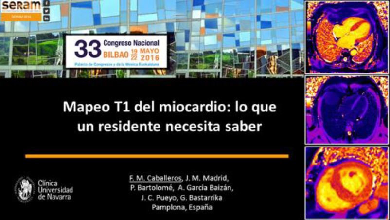
Mapeo T1 del miocardio:
lo que un residente necesita saber
Palabras clave:
Mapeo T1, miocardio, cardioresonancia magnéticaResumen
Objetivos Docentes
1. Revisar la fisiopatología de la fibrosis miocárdica.
2. Entender como funcionan las secuencias de mapeo T1.
3. Enfatizar la relevancia científica y clínica del mapeo T1.
Revisión del tema
INTRODUCCION
La cardioresonancia magnética (CRM) se ha establecido como estándar de referencia para la evaluación de la anatomía y función cardíaca. Permite, además, caracterizar el tejido miocárdico de manera no invasiva. Habitualmente esto se realiza tras la administración de contraste paramagnético y mediante la técnica de realce tardío. De esta manera, es posible detectar cambios macroscópicos y clasificar muchas miocardiopatías. Sin embargo, los cambios más difusos, en los que no existe una diferencia significativa en intensidad de señal entre el miocardio sano y enfermo, no se pueden detectar.
Para este fin se han desarrollado nuevas secuencias basadas en técnicas paramétricas, entre las que se incluyen el mapeo T1.
Descargas
Citas
Moon, J., Messroghli, D., et al. Myocardial T1 mapping and ECV quantification. JCMR. 2013, 15:92
Perea Palazon, R, OrtizPerez, J. et al. Characterising the myocardial interstitial space: clinical relevance of T1 and extracellular volume mapping. ECR: DOI: 10.1594/ecr2014/C0416
Scot, A., Travis, S. et al Mapping the Future of Cardiac MR Imaging: Casebased Review of T1 and T2 Mapping Techniques. RadioGraphics 2014; 34:1594–1611
Messroghli,D., Radjenovic,A., et al. MOLLI for high resolution T1 mapping of the heart. Magnetic Resonance in Medicine 2004, 52:141–146.
Kellman, P., Hansen, M. T1 Mapping in the heart: accurancy and precision. JCMR. 2014, 16:2
Jellis, C., Kwon.H. Myocardial T1 mapping: modalities and clinical applications. Cardiovasc Diagn Ther 2014;4(2):126137.
Piechnik SK et al. Shortened Modified LookLocker Inversion recovery (ShMOLLI) for clinical myocardial T1mapping at 1.5 and 3 T within a 9 heartbeat breathhold. J Cardiovasc Magn Reson 2010;19;12:69
Lee JJ et al. Myocardial T1 and Extracellular Volume Fraction Mapping at 3 Tesla. Journal of Cardiovascular Magnetic Resonance 2011, 13:75 52.
Klein CT et al. The influence of myocardial blood flow and volume of distribution on late GdDTPA kinetics in ischemic heart failure. J Magn Reson Imaging. 2004;20:58893. 53. Klein C et al.
Rogers TJ et al. Standardization of T1 measurements with MOLLI in differentiation between health and disease the ConSept study. Cardiovasc Magn Reson. 2013;15:78 55.
Von KnobelsdorffBrenkenhoff F et al. Myocardial T1 and T2 mapping at 3 T: reference values, influencing factors and implications J Cardiovasc Magn Reson. 2013;15:53
Hansen, M., Merchant, N. MRI of Hypertrophic Cardiomyopathy: Part I, MRI Appearances. AJR 2007, 189: 13351343.
Parsai, C. et al. Diagnostic and prognostic value of cardiovascular magnetic resonance in nonischaemic
cardiomyopathies. Journal of Cardiovascular Magnetic Resonance 2012, 14:54
Karamitsos TD, Piechnik SK, Banypersad SM, et al. Noncontrast T1 mapping for the diagnosis of cardiac amyloidosis. JACC Cardiovasc Imaging 2013;6(4): 488–497.
Fontana M, Banypersad SM, Treibel TA, et al. Native T1 mapping in transthyretin amyloidosis. JACC Cardiovasc Imaging 2014;7(2):157–165.
Ugander M, Bagi PS, Oki AJ, et al. Myocardial edema as detected by precontrast T1 and T2 CMR delineates area at risk associated with acute myocardial infarction. JACC Cardiovasc Imaging 2012; 5 (6):596–603.
Bruder O, Wagner A, Jensen CJ, et al. Myocardial scar visualized by cardiovascular magnetic resonance imaging predicts major adverse events in patients with hypertrophic cardiomyopathy. J Am Coll Cardiol 2010;56 (11):875–887.
Sibley CT, Noureldin RA, Gai N, et al. T1 mapping in cardiomyopathy at cardiac MR: comparison with endomyocardial biopsy. Radiology 2012;265(3):724–732.


