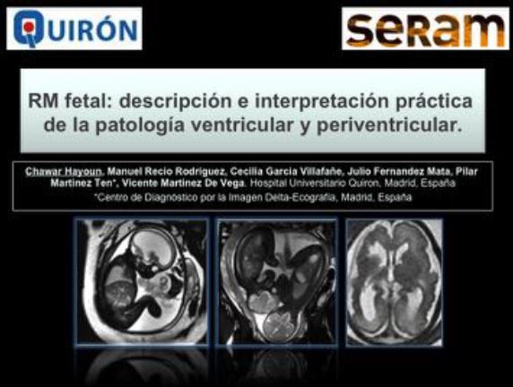
RM fetal:
descripción e interpretación práctica de la patología ventricular y periventricular.
Palabras clave:
patología ventricular, poster, seram, RM fetal, patología periventricularResumen
Objetivos Docentes
Revisar e ilustrar con ejemplos, las manifestaciones en resonancia magnética (RM) de las alteraciones ventriculares y periventriculares del cerebro fetal.
Integrar con una visión global, los hallazgos radiológicos para el diagnóstico de las distintas patologías.
Revisión del tema
INTRODUCCIÓN
La ecografía (US) es la técnica de imagen de elección para el examen de rutina del sistema nervioso central (SNC) fetal, pero sus hallazgos son frecuentemente inespecíficos y tiene varias limitaciones técnicas, que disminuyen su ya de por si baja sensibilidad.
La resonancia magnética (RM) como prueba complementaria, tiene varias ventajas en el estudio del cerebro fetal. Por su valoración volumétrica y su mayor contraste entre tejidos, permite la visualización al mismo tiempo de ambos hemisferios cerebrales, y una mejor evaluación del desarrollo cortical y del patrón de sulcación cerebral.
Descargas
Citas
•Manuel Recio Rodriguez, Vicente Martinez de Vega Fenández, Pilar Martinez Ten, Javier Perez Pedregosa, Daniel Martin Fernández-Mayorales, Mar Jimenez de la Peña. RM fetal en las anomalías del SNC. Aspectos de interés para el obstetra. RAR 2010.
•Glenn OA, Barkovich AJ. Magnetic Resonance Imaging of he Fetal Brain and Spine: An Increasingly Important Tool in Prenatal Diagnosis. AJNR Am J Neuroradiol 2006
•Garel C. The role of MRI in the evaluation of the fetal brain with an emphasis on biometry, gyration and parenchyma. Pediatr Radiol 2004
•Laurent Guibaud. Contribution of fetal cerebral MRI for diagnosis of structural anomalies. Prenatal diagnosis 2009.
•Garel C, Alberti C. Coronal measurement of the fetal lateral ventricles: comparison between ultrasonography and mag- netic resonance imaging. Ultrasound Obstet Gynecol 2006
•Deborah Levine, MD Patrick D. Barnes, MD, Joseph R. Madsen. Fetal central nervous system anomalies: MR imaging augment sonographic diagnosis. Radiology 1997.
•Steven W. Hetts Elliott H. Sherr Stephanie Chao Sarah Gobuty A. James Barkovich.. Anomalies of the Corpus Callosum: An MR Analysis of the Phenotypic Spectrum of Associated Malformations. AJR 2006
•Catherine Garel, Anne-Lise Delezoide, Monique Elmaleh-Berges, Fran ¸coise Menez, Catherine Fallet-Bianco, Edith Vuillard, Dominique Luton, Jean-Fran ¸cois Oury, and Guy Sebag. Contribution of Fetal MR Imaging in the Evaluation of Cerebral Ischemic Lesions. AJNR 2004
•Abdel Razek AA, Kandell AY, Elsorogy LG., Elmongy A, Basett AA. Disorders of cortical formation: MR imaging fea- tures. AJNR Am J Neuroradiol 2009
•Glenn OA, Goldstein R, Li KC, et al. Fetal MRI in the evaluation of fetuses referred for sonographically suspected abnor- malities of the corpus callosum. J Ultrasound Med 2005
•Garel C, Delezoide AL, Elmaleh-Berges M, et al. Contribution of fetal MR imaging in the evaluation of cere- bral ischemic lesions. AJNR Am J Neuroradiol 2004
•Benoist G, Salomon LJ, Mohlo M, Suarez B, Jacqucmard F, Ville Y. Cytomegalovirus-related fetal brain lesions: compa- rison between targeted ultrasound examination and magnetic resonance imaging. Ultrasound Obstet Gynecol 2008.


