Manifestaciones neurorradiológicas en el síndrome de embolia grasa.
Palabras clave:
poster, seram, embolia grasa, EG, SEGResumen
Objetivos Docentes
Revisar el síndrome de embolia grasa, con énfasis en los hallazgos neurorradiológicos.
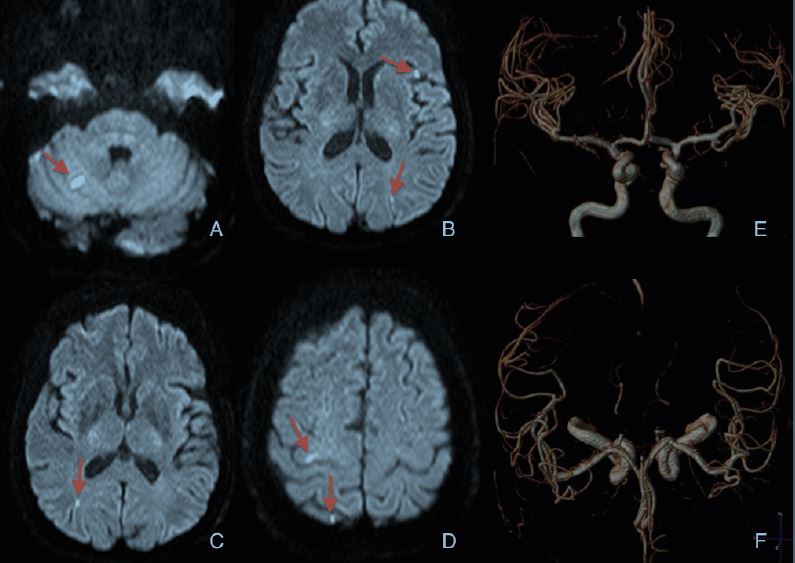
Revisión del tema
La embolia grasa (EG) se define por la presencia de partículas de grasa en la circulación sanguínea. Fue descrita por
primera vez por Zenker en 1861, siendo un evento común reportado hasta en un 90% de pacientes politraumatizados, que por
lo general cursa de forma asintomática. Cuando presenta manifestaciones clínicas se denomina síndrome de embolia grasa
(SEG), con una incidencia que oscila según autores entre 1% - 30% (amplio rango que probablemente refleja la
heterogeneidad clínica y de criterios diagnósticos).
Descargas
Citas
K.-H. Kuo, Y.-J. Pan, Y.-J. Lai, et-al. Dynamic MR Imaging Patterns of Cerebral Fat Embolism: A Systematic Review with Illustrative Cases AJNR Am J Neuroradiol 2014 35: 1052-1057.
Zaitsu Y, Terae S, Kudo K et-al. Susceptibility-weighted imaging of cerebral fat embolism. J Comput Assist Tomogr. 2010;34 (1): 107-12.
Butteriss D, Mahad D, Soh C, Walls T, Weir D, Birchall D. Case report. Reversible cytotoxic cerebral edema in cerebral fat embolism AJNR Am J Neuroradiol 2006, 27:620-3.
Shaikh N, Parchani A, Bhat V et-al. Fat embolism syndrome: clinical and imaging considerations: case report and review of literature. Indian J Crit Care Med. 2008;12 (1): 32-6.
Chrysikopoulos H, Maniatis V, Pappas J et-al. Case report: post-traumatic cerebral fat embolism: CT and MR findings. Report of two cases and review of the literature. Clin Radiol. 1996;51 (10): 728-32.
Stoeger A, Daniaux M, Felber S et-al. MRI findings in cerebral fat embolism. Eur Radiol. 1999;8 (9): 1590-3. 7. Kawano Y, Ochi M, Hayashi K et-al. Magnetic resonance imaging of cerebral fat embolism. Neuroradiology. 1991;33 (1): 72-4.
Suh SI, Seol HY, Seo WK et-al. Cerebral fat embolism: susceptibility-weighted magnetic resonance imaging. Arch. Neurol. 2009;66 (9): 1170.
Kim H, Lee JH, Lee CH, et al. Experimental cerebral fat embolism: embolic effects of triolein and oleic acid depicted by MR imaging and electron microscopy. AJNR Am J Neuroradiol 2002; 23:1516-23.
Kim HJ, Lee CH, Kim HG, et al. Reversible MR changes in the cat brain alter cerebral fat embolism induced by triolein emulsion. AJNR 2004; 25: 958-63.
Parizel P, Demey H, Veeckmans G, et al. Early diagnosis of cerebral fat embolism syndrome by diffusion-weighted MRI (starfield pattern). Stroke 2001; 32:2942-4.