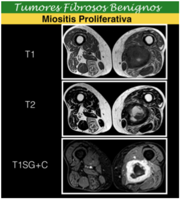
Tumores Fibroblásticos/Miofibroblásticos de los Tejidos Blandos: Orientación Según sus Hallazgos Radiológicos.
Palabras clave:
lesiones, tumores, Fibroblásticos/Miofibroblásticos, tejidos blandosResumen
-
Describir la gama de lesiones tumorales Fibroblásticos/Miofibroblásticos de los tejidos blandos según la nueva clasificación los tumores de partes blandas editada por la Organización Mundial de la salud (OMS) en el año 2013.
-
Mostrar los protocolos de adquisición de imágenes mediante Resonancia Magnética (RM) que se deben llevar a cabo para su diagnóstico.
-
Exponer los hallazgos radiológicos en RM de estas lesiones organizadas en los 4 subgrupos adoptados por la OMS, según su comportamiento biológico.
Descargas
Citas
Fox MG, Kransdorf MJ, Bancroft LW, Peterson JJ, Flemming DJ: MR imaging of fibroma of the tendon sheath. AJR American journal of roentgenology 2003, 180(5):1449-1453.
Lourie JA, Lwin KY, Woods CG: Case report 734. Fibroma of tendon sheath eroding 3rd metatarsal bone. Skeletal radiology 1992, 21(4):273-275.
Southwick GJ, Karamoskos P: Fibroma of tendon sheath with bone involvement. J Hand Surg
Br 1990, 15(3):373-375.
Greene TL, Strickland JW: Fibroma of tendon sheath. J Hand Surg Am 1984, 9(5):758-760
Beggs I, Salter DS, Dorfman HD: Synovial desmoplastic fibroblastoma of hip joint with bone
erosion. Skeletal radiology 1999, 28(7):402-406.
Shuto R, Kiyosue H, Hori Y, Miyake H, Kawano K, Mori H: CT and MR imaging of desmoplastic fibroblastoma. Eur Radiol 2002, 12(10):2474-2476.
Walker KR, Bui-Mansfield LT, Gering SA, Ranlett RD: Collagenous fibroma (desmoplastic fibroblastoma) of the shoulder. AJR American journal of roentgenology 2004, 183(6):1766.
Lee JC, Thomas JM, Phillips S, Fisher C, Moskovic E: Aggressive fibromatosis: MRI features with pathologic correlation. AJR American journal of roentgenology 2006, 186(1):247-254.
Masciocchi C, Lanni G, Conti L, Conchiglia A, Fascetti E, Flamini S, Coletti G, Barile A: Soft-tissue inflammatory myofibroblastic tumors (IMTs) of the limbs: potential and limits of diagnostic
imaging. Skeletal radiology 2012, 41(6):643-649.
Murphey MD, Ruble CM, Tyszko SM, Zbojniewicz AM, Potter BK, Miettinen M: From the archives of the AFIP: musculoskeletal fibromatoses: radiologic-pathologic
correlation. Radiographics2009, 29(7):2143-2173.
Murphey MD, Fairbairn KJ, Parman LM, Baxter KG, Parsa MB, Smith WS: From the archives of the AFIP. Musculoskeletal angiomatous lesions: radiologic-pathologic
correlation. Radiographics1995, 15(4):893-917.
Disler DG, Alexander AA, Mankin HJ, O'Connell JX, Rosenberg AE, Rosenthal DI: Multicentric fibromatosis with metaphyseal dysplasia. Radiology 1993, 187(2):489-492.
Romero JA, Kim EE, Kim CG, Chung WK, Isiklar I: Different biologic features of desmoid tumors in adult and juvenile patients: MR demonstration. J Comput Assist Tomogr 1995,19(5):782-787.
Dinauer PA, Brixey CJ, Moncur JT, Fanburg-Smith JC, Murphey MD: Pathologic and MR Imaging Features of Benign Fibrous Soft-Tissue Tumors in Adults1. Radiographics 2007, 27(1):173-187.
Taylor LJ: Musculoaponeurotic fibromatosis. A report of 28 cases and review of the literature. Clin Orthop Relat Res 1987(224):294-302.
Enzinger FM, Smith BH: Hemangiopericytoma. An analysis of 106 cases. Hum
Pathol 1976,7(1):61-82.


